Reading radiographic images in chronological order offers greater sensitivity for detecting disease progression in osteoarthritis (OA), according to research presented at the 2005 European Congress of Rheumatology in Vienna, Austria.
Dr. Margreet Kloppenburg and colleagues enrolled 20 patients with osteoarthritis in their two-year study to compare chronological radiograph reading to reading in paired x-rays. Kloppenburg's group is from the department of clinical epidemiology and rheumatology at Leiden University Medical Center in the Netherlands.
The patients had osteoarthritis in at least two joints, and were imaged at baseline and again at two years. Two radiologists read the x-rays and scored them in consensus. They graded joint space narrowing and osteophytes on a 0-3 scale. Additionally, they graded the cervical (C2-7) and lumbar spine disk spaces (L1-S1) in the same manner (0-3) looking for disk space narrowing and anterior osteophytes using the Lane atlas.
Radiologists read all the films in pairs, with an unknown time sequence, and in chronological order. Radiological progression was defined as an increase of at least one grade in joint space narrowing, disk space narrowing, or osteophyte total scores in the different joint groups. Researchers then compared the two procedures utilizing the standardized response mean (SRM).
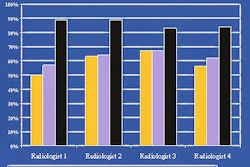
Kloppenburg reported that paired reading showed progression of joint or disk space narrowing scores in the hands in 5% of patients, hips in 5%, knees in 20%, and spine in 20%.
However, progression was more evident with chronological reading of images. Joint or disk space narrowing progression was visible in the hands in 15% of patients, hips in 10%, knees in 25%, and spine in 20%.
In terms of osteophyte scores, paired readings revealed progression of osteophyte scores in the hands (15%), hips (10%), knees (10%), and spine (25%). Chronological readings showed a progression in patients' hands (15%), hips (10%), knees (25%), and spine (35%).
Finally, Kloppenburg noted that the SRM for changes in joint or disk space narrowing progression scores based on paired readings was 0.00 in both the hands and hips, 0.32 in the knees, and 0.13 in the spine. For chronological readings, the SRM for change was 0.38 in the hands, 0.32 in the hips, 0.56 in the knees, and 0.18 in the spine. The investigators said they found similar levels of sensitivity in the SRM for changes in osteophyte progression scores.
Although the patient population in this study was small, Kloppenburg concluded that the findings still had implications for the choice of reading procedure in progression studies for patients with osteoarthritis.
By Jerry Ingram
AuntMinnie.com contributing writer
June 30, 2005
Related Reading
MRI, x-ray yield comparable sensitivities for RA joint erosion, June 10, 2005
Tibial-talar ratio on x-ray reliably shows ankle alignment, May 6, 2005
Copyright © 2005 AuntMinnie.com