AuntMinnie
  • Europe
  • Clinical News
  • Informatics
  • Industry News
  • Practice Management
  • Education
  • Subspecialties
  • More
  • Sign In
  • Board Review
  • CME
  • Careers
  • Cases
  • Conferences
  • Videos
  • Webinars
  • Podcast Network
  • Advertising
  • Buyer's Guide
  • Vendors
  • Minnies
  • RPT Tracker
Clinical News
  • CT
  • Digital X-Ray
  • Interventional
  • Molecular Imaging
  • MRI
  • Radiation Oncology/Therapy
  • Ultrasound
  • Womens Imaging
Imaging Informatics
  • Advanced Visualization
  • Enterprise Imaging
  • AI
  • Cybersecurity
Industry News
  • Installations
  • Product News
  • Regulatory
  • M&A
  • Market Analysis
Practice Management
  • Administration
  • Associations
  • Careers
  • Equity & Inclusion
  • Legislation
  • Medicolegal
  • Patient Safety
  • Radiologic Technologist
  • Service
  • Salary Scan
Resources
  • Board Review
  • CME
  • Careers
  • Cases
  • Conferences
  • Videos
  • Webinars
  • Podcast Network
  • Advertising
  • Buyer's Guide
  • Vendors
  • Minnies
  • RPT Tracker
User Tools
  • Sign In
More
  • About Us
  • Advertising
  • Help
Follow AuntMinnie
Facebook iconTwitter X icon LinkedIn iconInstagram icon
  • Europe
  • Clinical News
    • CT
    • Digital X-Ray
    • Interventional
    • Molecular Imaging
    • MRI
    • Radiation Oncology/Therapy
    • Ultrasound
    • Womens Imaging
  • Informatics
    • Advanced Visualization
    • Enterprise Imaging
    • AI
    • Cybersecurity
  • Industry News
    • Installations
    • Product News
    • Regulatory
    • M&A
    • Market Analysis
  • Practice Management
    • Administration
    • Associations
    • Careers
    • Equity & Inclusion
    • Legislation
    • Medicolegal
    • Patient Safety
    • Radiologic Technologist
    • Service
    • Salary Scan
  • Education
    • Medical Students
    • Residents/Fellows
  • Subspecialties
    • Breast Imaging
    • CV
    • Chest
    • Emergency
    • GI
    • GU
    • Head & Neck
    • Interventional
    • Physics
    • MSK
    • Neuro
    • Nuclear
    • Pediatric
    • Radiation Oncology
  • More
  • Board Review
  • CME
  • Careers
  • Cases
  • Conferences
  • Videos
  • Webinars
  • Podcast Network
  • Advertising
  • Buyer's Guide
  • Vendors
  • Minnies
  • RPT Tracker
  • Sign In
Follow AuntMinnie
Facebook iconTwitter X icon LinkedIn iconInstagram icon

Top Story

Ultrasound Portable 400
Ultrasound
POCUS improves timeliness in emergency pregnancy care
Emergency POCUS can reduce length of stay and time to diagnosis while not leading to more missed diagnoses of ectopic pregnancy.
Featured
This figure shows fused I-124 evuzamitide positron emission tomography (PET)/computed tomography (CT) images from patients without amyloidosis and from patients with ATTR cardiac amyloidosis, early ATTR cardiac amyloidosis, light chain cardiac amyloidosis, gelsolin-mediated amyloidosis, and systemic organ amyloid. The columns represent radiological transaxial, sagittal, and coronal views except for the last row middle and far right columns, which are coronal projections. The blue asterisks represent physiological activity (tracer activity in the stomach and esophagus). The arrowheads represent pathological uptake. Note the small amount of cardiac uptake in the basal septum and the basal lateral walls (likely early ATTR cardiac amyloidosis), splenic uptake and pleural uptake in light chain amyloidosis, and cardiac and kidney uptake in gelsolin-mediated amyloidosis. The bright yellow color represents more tracer uptake.
Molecular Imaging
Bayer's PET agent meets endpoints in phase III cardiac amyloidosis study
Brain 400
MRI
MRI-based biomarker tool can predict progression to dementia
Latest News
Representative case from the non-nitroglycerin cohort. A 92-year-old man who did not receive sublingual nitroglycerin because of severe aortic stenosis. The Agatston coronary artery calcium score was 5438. (A) Curved multiplanar reformatted CCTA image reconstructed at 0.4-mm slice thickness using a Bv60 kernel and QIR strength 3 demonstrates diffuse multifocal mild luminal stenoses related to extensive calcified atherosclerotic plaque. (B) Invasive coronary angiography confirmed multifocal mild stenoses without evidence of obstructive coronary artery disease.
Is nitroglycerin necessary prior to photon-counting CT?
August 27, 2026
Comparison of contrast-enhanced CT (ceCT) and FAPI-PET in detecting peritoneal carcinomatosis in patient with advanced ovarian carcinoma. (A) ceCT shows subtle peritoneal thickening that is difficult to delineate. (B) Maximum-intensity-projection image demonstrating diffuse intense FAPI uptake corresponding to extensive peritoneal carcinomatosis. (C) FAPI-PET shows intense uptake corresponding with peritoneal carcinomatosis, with improved lesion conspicuity over ceCT.
PET outperforms CT in ovarian cancer
August 28, 2026
Examples of ultrasound findings and techniques. (A) Images in a 39-year-old male patient with a mass in the left thigh. The mass is heterogeneous on the B-mode US image (compared with the patient in D) and showed increased microvascularity (superb microvascular imaging [SMI]) and shear-wave elastography (SWE) values. Undifferentiated pleomorphic sarcoma was diagnosed at biopsy (with pleomorphic rhabdomyosarcoma in surgical specimen). (B) Images in an 18-year-old male patient with a mass in the left leg. The mass is hypoechoic on the B-mode image, with no other findings suggestive of malignancy. The lesion is in contact with the cortex of the tibia, which is slightly irregular. CT revealed a doubtful anteromedial tibial erosion. The microvascular study demonstrated high vascularization, suggestive of malignancy. Periosteal Ewing sarcoma was diagnosed with both histologic and immunohistochemical confirmation. (C) Images in a 69-year-old female patient with a lump growing on the outside of the left leg. Multiple SWE examinations were performed (please note the high values obtained in the measurements, whereas the color map highlights the stiffness relative to adjacent tissues). SMI showed areas of increased vascularization to target for sampling. Undifferentiated spindle cell sarcoma was diagnosed at biopsy, with residual leiomyosarcoma in the surgical specimen after neoadjuvant therapy. (D) Images in a 56-year-old female patient with a mass in the right thigh. The mass is heterogeneous at both B-mode ultrasound (similar to patient A) and MRI (coronal T2-weighted spectral attenuated inversion recovery [SPAIR]; T1-weighted pre-contrast and postcontrast imaging), which even shows uptake after the administration of paramagnetic contrast material, which is traditionally suggestive of malignancy. Low values at SMI and elastography are suggestive of benignity. Spindle cell lipoma was diagnosed at biopsy, with atypical spindle cell lipomatous tumor in the surgical specimen.
Advanced ultrasound techniques improve soft tissue tumor evaluation
August 26, 2026
Choropleth map of state-level imaging-associated greenhouse gas emissions per 1,000 Medicare Part B fee-for-service beneficiaries in 2022. Darker shades indicate higher per-beneficiary greenhouse gas emissions. District of Columbia (not shown) is in 3-4 MTCO2e per 1,000 range. MT COâ‚‚e = metric tons of carbon dioxide equivalent
Sustainability efforts should look to state power grids
August 28, 2026
Podcast Network
Latest Episode
Podcast: Where do MRI accident reports go?
More episodes »
Cases of the Week
Check out our Cases of the Week!
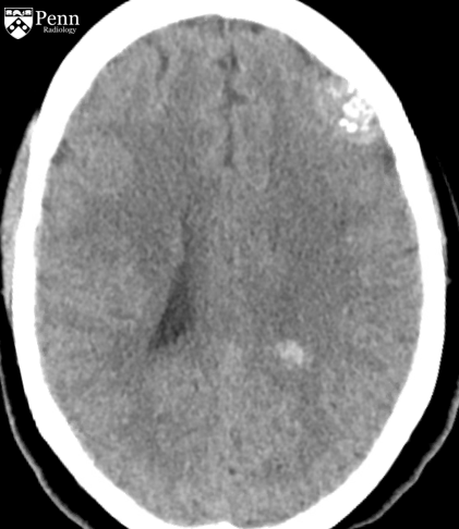
Patient with abdominal pain revisited
A patient with abdominal pain out of proportion to the exam.
39-year-old man with chronic abdominal pain revisited
83-year-old female former smoker with imaging follow-up revisited
View All Cases
Clinical News
This figure shows fused I-124 evuzamitide positron emission tomography (PET)/computed tomography (CT) images from patients without amyloidosis and from patients with ATTR cardiac amyloidosis, early ATTR cardiac amyloidosis, light chain cardiac amyloidosis, gelsolin-mediated amyloidosis, and systemic organ amyloid. The columns represent radiological transaxial, sagittal, and coronal views except for the last row middle and far right columns, which are coronal projections. The blue asterisks represent physiological activity (tracer activity in the stomach and esophagus). The arrowheads represent pathological uptake. Note the small amount of cardiac uptake in the basal septum and the basal lateral walls (likely early ATTR cardiac amyloidosis), splenic uptake and pleural uptake in light chain amyloidosis, and cardiac and kidney uptake in gelsolin-mediated amyloidosis. The bright yellow color represents more tracer uptake.
Molecular Imaging
Bayer's PET agent meets endpoints in phase III cardiac amyloidosis study
MRI
MRI-based biomarker tool can predict progression to dementia
CT
Is nitroglycerin necessary prior to photon-counting CT?
Molecular Imaging
PET outperforms CT in ovarian cancer
Ultrasound
Advanced ultrasound techniques improve soft tissue tumor evaluation
View More »
AI
Artificial Intelligence Ai Global Communication Network 400
AI
Most FDA-cleared AI medical devices not tested on patient outcomes
MRI
AI framework could improve MRI quality control in prostate cancer
Womens Imaging
Longitudinal DBT could help improve long-term cancer risk prediction
AI
AI tools may not meet breast radiologists' expectations in practice
AI
Model confidence, reader expertise affect reader-LLM collaboration
View More »
Industry News
Cash
Enterprise Imaging
Scan.com raises $220M
PACS/VNA
Novarad releases NovaPACS update
Ultrasound
GEHC adds three systems to Vivid ultrasound portfolio
Practice Management
Mid-size radiology practices may lose $2.6M annually to billing gaps
AI
Nanox AI signs distribution deal for HealthOST software
View More »
Practice Management
Acr2
ACR Foundation receives $6M gift
The funds will support public policy research, data analysis, and regulatory policy efforts across the ACR.
Sustainability efforts should look to state power grids
Mid-size radiology practices may lose $2.6M annually to billing gaps
More Practice Management
More from AuntMinnie
Scan.com raises $220M
By AuntMinnie.com staff writers
The company will use the funds to invest in AI and application programming interface (API) infrastructure for patient scheduling and diagnostics.
August 31, 2026
Cash
ACR Foundation receives $6M gift
By AuntMinnie.com staff writers
The funds will support public policy research, data analysis, and regulatory policy efforts across the ACR.
August 31, 2026
Acr2
Novarad releases NovaPACS update
By AuntMinnie.com staff writers
Version 9.1 is a a major security upgrade.
August 28, 2026
Pacs
GEHC adds three systems to Vivid ultrasound portfolio
By AuntMinnie.com staff writers
The company will showcase the ultrasound systems at the European Society of Cardiology (ESC) Congress 2026 in Munich.
August 28, 2026
From left to right: Vivid Explorer, Vivid Advanced, and Vivid Focus
Women's Imaging MinnieCast, Episode 16: Breast AI expectations versus performance
By Amerigo Allegretto
Azadeh Elmi, MD, shares results showing how breast imaging AI tools have modest clinical benefit in practice over expectations.
August 28, 2026
Minnie Cast Thumbnail (21)
Mid-size radiology practices may lose $2.6M annually to billing gaps
By AuntMinnie.com staff writers
Reducing denials and underpayments, operational efficiencies, and patient collections may be contributors.
August 27, 2026
Business Financial Results 400
Nanox AI signs distribution deal for HealthOST software
By AuntMinnie.com staff writers
The distribution deal with Vertec Scientific covers an initial three-year term.
August 27, 2026
Business Handshake 400
CT reveals progressive growth pattern in incidental lung nodules
By Will Morton
This pattern is most associated with a family history of lung cancer.
August 25, 2026
Serial axial chest CT scans demonstrating the longitudinal evolution of small pure ground-glass nodules (pGGNs) incidentally detected during a routine wellness assessment. (A) Images in a 37-year-old woman with a family history of lung cancer show a nodule with the progressive growth pattern (box). The baseline CT scan, obtained in October 2017, demonstrates a pure ground-glass nodules in the right lower lobe. Consecutive CT scans obtained from 2017 to 2022 show the continuous enlargement of the nodule during active surveillance. The maximum diameter of the nodule increased from 5.6 mm in 2017 to 10.2 mm in 2022. (B) Images in a 45-year-old man show a nodule with the indolent growth pattern (box). The baseline CT scan, obtained in April 2018, demonstrates a pure ground-glass nodules in the left lower lobe. Consecutive CT scans obtained from 2019 to 2023 show that the nodule remained radiographically stable during CT surveillance. The maximum diameter of the nodule was 5.4 mm in 2018 and 6.1 mm at the final surveillance scan in 2023.
Should cancer data stop lumping Asian Americans, NHPI people together?
By Amerigo Allegretto
The results highlight the need for disaggregated data to effectively target cancer prevention and control interventions.
August 27, 2026
Woman Patient Asian 400
Radiology faculty most stressed, least satisfied
By Will Morton
Across faculty, technologists, and nurses, the most common stressors were generally system-level issues.
August 27, 2026
Shutterstock 1865403334
Radiology Partners to acquire Everlight Radiology in $1 billion deal
By AuntMinnie.com staff writers
Radiology Partners said it intends to make its Mosaic Drafting AI tool available to Everlight radiologists.
August 25, 2026
Acquisitions
Philips, Imricor launch MRI-guided cardiac intervention lab platform
By AuntMinnie.com staff writers
The platform combines Philips' 1.5T MRI system with Imricor's MR-compatible systems for MR-guided cardiac ablation.
August 27, 2026
Philips Rsna 2021 400
AuntMinnie
Topics
  • Clinical News
  • Imaging Informatics
  • Industry News
  • Practice Management
  • Radiology Education
  • Subspecialties
Resources
  • Board Review
  • CME
  • Careers
  • Cases
  • Conferences
  • Videos
  • Webinars
  • Podcast Network
  • Advertising
  • Buyer's Guide
  • Vendors
  • Minnies
  • RPT Tracker
More
  • About Us
  • Advertising
  • Help
Follow AuntMinnie
Facebook iconTwitter X icon LinkedIn iconInstagram icon
  1. Privacy Policy
  2. Privacy Settings
  3. Terms & Conditions
  4. Contact Us
  5. Site Map
  6. Home
© 2026 Science and Medicine Group. All rights reserved.