Mesenchymal Hamartoma of the Chest Wall:
Clinical:
Mesenchymal hamartomas of the chest wall are rare lesions (about 60 reported cases) of early infancy and childhood [1]. The lesions always arises in a rib and constitutes a benign proliferation of skeletal tissue with a prominent cartilagenous component and hemorrhagic cavities (secondary aneurysmal bone cyst formation) [1]. The lesion has no potential for invasion or metastases [1]. The most common finding is a deforming chest wall mass noted at birth [1].
X-ray:
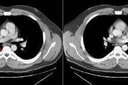
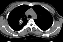
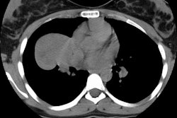
The typical finding is a large, extrapleural, partially calcified soft-tissue mass arising from one or more ribs with associated destruction and distortion of the adjacent osseous thorax [1]. The appearance suggests an aggressive lesion, but this is not the case [1]. CT will demonstrate a large, heterogeneous mass containing a calcified or chondroid matrix [1]. Hemorrhagic cystic areas with fluid levels are seen in about 65% of cases on CT and in 80% of cases on MR [1].
REFERENCES:
(1) Radiology 2001; Groom KR, et al. Mesenchymal hamartoma of the chest wall: radiologic manifestations with emphysis on cross-sectional imaging and histopathologic comparison. 222: 205-211


