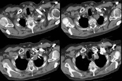
Pulmonary Hamartoma
The patient shown in the case below was referred for evaluation of a right upper lobe mass. The CT scan demonstrates a large mass in the right upper lobe that contained macroscopic fat consistent with a hamartoma. Lung windows demonstrated the lesion to be endobronchial and this was confirmed at surgical resection. (Click images to enlarge)