Sclerosing hemangioma:
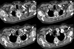
The images demonstrate an enhancing soft tissue mass within the right lung. The mass has areas of slightly decreased attenuation best appreciated following IV contrast administration. (Click images to enlarge)
Non-contrast CT:

Post contrast CT with enhancement and areas of decreased attenuation:

CT lung windows reveals the mass to lie along the minor fissure- this was confirmed at surgery:

T2 weighted MR images demonstrate a region of increased signal intensity within the mass suggesting necrosis:

Post gadolinium images demonstrate the mass to enhance relatively homogeneously, but note that the area of necrosis does not enhance: