- Radiology 2000 Jul;216(1):117-21
- Pulmonary tuberculoma evaluated by means of FDG PET: findings in 10
cases.
Goo JM, Im JG, Do KH, Yeo JS, Seo JB, Kim HY, Chung JK
Department of Radiology and Institute of Radiation Medicine, Seoul National University Hospital, 28, Yongon-Dong, Chongno-Gu, Seoul, 110-744, South Korea.
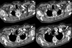
PURPOSE: To describe findings of pulmonary tuberculoma at 2-[fluorine 18]fluoro-2-deoxy-D-glucose (FDG) positron emission tomography (PET). MATERIALS AND METHODS: Ten consecutive patients who underwent PET and subsequently were proved to have pulmonary tuberculoma were analyzed. Tuberculosis was proved histopathologically in eight by means of wedge resection or lobectomy (n = 7) or needle biopsy (n = 1) and in two by means of clinical follow-up for more than 2 years. PET scans were evaluated by using peak standardized uptake values. Computed tomographic (CT) and histopathologic findings also were reviewed. RESULTS: Nine of 10 tuberculomas showed FDG uptake at PET, and the mean peak standardized uptake value was 4.2 +/- 2.2 (SD). FDG uptake (range, 1. 9-3.7) in lesions adjacent to main abnormalities was demonstrated in four patients. On CT scans, the mean of the longest nodule diameters was 21 mm +/- 8, and there were some areas of branching linear opacities or satellite nodules that suggested pulmonary tuberculosis in seven patients. Histopathologic findings were chronic granulomatous inflammation with caseation necrosis (n = 7) and healed tuberculosis with aspergilloma (n = 1). CONCLUSION: Pulmonary tuberculoma commonly causes an increase in FDG uptake. These results suggest that in geographic regions with a high prevalence of granulomatous lesions, positive FDG PET results should be interpreted with caution in differentiating benign from malignant pulmonary abnormalities.
PMID: 10887236, UI: 20347960
Tumor > Malignant > Lungcancer
Latest in Tumor
Tumor > Benign > Inflammatory myofibroblastic tumor
October 19, 2020
Tumor > Malignant > Lungcancer > Staging
February 22, 2009
Tumor > Benign > Mesenchymal hamartoma
October 30, 2008
Tumor > Benign > Leiomyoma
October 30, 2008