Leiomyoma:
Clinical:
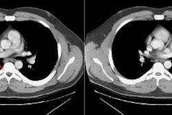
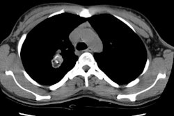
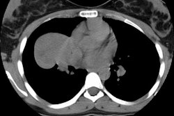
Pulmonary leiomyoma is a rare tumor [1]. The lesion is located within the bronchi in 45-66% of patients, the trachea in up to 1/3 of patients, and the remainder are found in the lung parenchyma [1] (other authors suggest the tracha is the most common site accounting for 2/3's of tumors [2]). Tracheal tumors originate from the smooth muscle in the tracheal wall- particualrly the posterior membranous portion [2]. The tumor is most common in the 4th decade of life, although one-third of patients are younger than 20 years [1]. The prognosis (excluding low grade leiomyosarcoma) is excellent following complete resection [1]. The presence of increased mitotic activity (greater than 3/10 high power fields), cytologic atypia, and necrosis are findings suggestive of leiomyosarcoma [1].
X-ray:
Most lesions appear as enhancing endoluminal masses [1]. An "iceberg" growth pattern is common- with a small intraluminal component and a large extraluminal component [1]. Calcification is rare [1].
REFERENCES:
(1) AJR 2007; Kim YK, et al. Airway leiomyoma: imaging findings and histopathologic comparisons in 13 patients. 189: 393-399
(2) Radiographics 2009; Park CM, et al. Tumors in the tracheobronchial tree: CT and FDG PET features. 29: 55-71


