Bone Imaging
Trauma:
Fractures: General findings
Normally, 80% of bone scans will show increased activity at a site of fracture by 24
hours, and 95% by 72 hours, although older (over 75 y) and debilitated patients may not
show activity for several days to as much as 2 weeks.
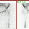
An illustrated report of an occult
metatarsal fracture is available at Washington University.
There are generally 3 phases identified when evaluating fractures:
There is increased flow and blood pool activity with diffuse and somewhat poorly defined increased delayed activity about the fracture site which is wider than the fracture line. Gallium uptake at acute fracture sites is usually increased as well. Mildly increased uptake of In-111 WBC's at the fracture site is present in up to 40% of acute uncomplicated fractures and may be associated with callus formation or adjacent myositis ossificans. Significant accumulation of white blood cells at the site of a healing fracture DOES NOT generally occur (seen in only 4% of cases) unless there is a complicating osteomyelitis.
Flow and blood pool abnormalities diminish considerably and delayed activity becomes more localized (focal at the fracture site) and intense.
A gradual decline in activity occurs over time with about 65% of exams normalizing by 1 year, and 90% becoming normal by 2 years. Persistent activity can be seen if healing has not produced normal anatomic alignment in weight bearing areas due to continued biomechanical stress and remodelling. Non-union occurs when there is failure of the fracture site to heal by 6 to 8 months following the injury. The tibia is the most frequent site of non-union. Non-union may occur secondary to an inadequate blood supply, osteoporosis, or infection. Two types of non-union have been described: reactive and atrophic.
Reactive Non-union:
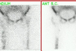
Characterized by persistent intense or normal uptake at the fracture site (80%). This bone has potential for healing (i.e.: evidence of metabolic activity) and these fractures tend to show significant improvement following electrical stimulation. Indium labeled WBC's are probably more reliable for detecting infection at the site of reactive non-united fractures, as gallium will also accumulate at these sites. It is not possible to distinguish a reactive non-union from a delayed union.
Atrophic Nonunion:
Appears as a photon deficient band between the fracture ends, which themselves may have more or less activity. This group of non-united fractures is usually not responsive to percutaneous electric stimulation.
Child Abuse:
Fractures of the femur or humerus, especially in children less than 1 years of age are very suspicious for child abuse. In the setting of the battered child, metaphyseal fractures near the growth plates, healed fractures, and skull fractures may be very difficult to visualize scintigraphically. It is best to perform a radiographic survey initially, and follow with scintigraphy if more information is required. Scintigraphy does have increased sensitivity for the detection of rib fractures compared to plain radiographs [1].
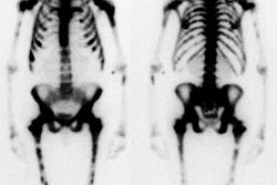
Vertebral Compression Fracture:
Regarding vertebral compression fractures, as with other fractures, most elderly patients will have a positive bone scan by 48 hours, however, the exam may not become positive for 3 to 7 days in up to 5-10% of patients. By 1 year, about 60% of compression fractures will normalize and 95% will become normal by 3 years. When multiple fractures are present in different stages of healing, osteoporosis is the most likely diagnosis. Multiple myeloma may occasionally mimic this appearance.
Stress/Insufficiency Fractures:
Stress fractures occur due to repetitive forces placed on normal bone. During
remodeling, there is a period when resorption exceeds replacement of bone which
temporarily weakens the cortex. Microfractures can result at these sites of resorption if
the stress is continually applied during this period. Gross fracture may result if these
microfractures are not permitted to heal. Insufficiency fractures result from normal or
physiologic loads placed on bone with deficient elastic resistance.
Radiographically, stress fractures appear as a lucent fracture line, focal sclerosis due
to endosteal callous formation, a periosteal reaction, or as an external callous. On bone
scan stress fractures usually appear as solitary, focal, fusiform areas of increased
tracer activity generally lasting for 10 to 12 weeks. Increased flow (for up to 3 to 4
weeks after onset of pain) and increased blood pool (for 6 to 8 weeks) activity may also
be seen. The scintigraphic abnormality often precedes the radiographic changes by 1 to 2
weeks. The metatarsals and the posterior medial cortex of the tibia are the most common
locations for stress fractures. Stress fractures of the tarsal navicular bone are
uncommon, but can produce foot pain in athletes. They most commonly involve the central
one third of the proximal dorsal margin of the tarsal navicular bone.
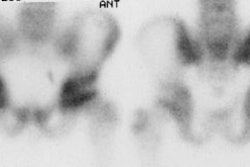
In the sacrum, stress fractures are associated with osteoporosis or prior radiation therapy to the pelvis (an insufficiency fracture). The osteoporotic sacrum develops a characteristic fracture with fracture lines running vertically through the left and right sides of the bone just medial to the SI joints, in conjunction with a transverse fracture just below the level of the SI joints. The fractures appear as a hot, geographic lesions confined to the sacrum and often have a characteristic "butterfly" or "Honda sign" appearance. Plain film are typically normal, but may show a sclerotic lesion in sacral ala.
Shin splints (stress periosteal reaction) are due to abnormal movement of the either the soleus muscle tendon complex or posterior tibialis muscle with disruption of Sharpey's fibers which produces a periostitis type lesion. Shin splints are characterized by abnormal linear (not focal) uptake of tracer along the posteromedial tibial cortex. A similar problem involving the tibialis anterior muscle has been suggested for abnormal activity along the anterior tibial cortex. Shin splints have not been demonstrated to progress to stress fractures.
Tendonitis may occasionally be detected on bone scan. Plantar fasciitis shows increased activity at the calcaneal tubercle. Patellar tendonitis at the inferior pole of the patella. Achilles tendonitis will show focal increased activity at the Achilles insertion on the posterior calcaneus (Differential diagnosis includes retrocalcaneal bursitis which usually appears more diffuse and less focal). Medial epicondylitis demonstrates focal increased activity over the medial humeral epicondyle at the insertion of the pronator flexor group.
Spondylolysis:
Spondylolysis is felt to represent a fatigue type fracture that occurs secondary to repetitive minor trauma. The sports most commonly associated with spondylolysis are gymnastics, diving, and contact sports such as football, hockey, and lacrosse. Approximately 90% of pars defects occur at the L5 level. With a unilateral pars defect (30% of cases), sclerosis, thickening, and enlargement of the contralateral lamina and pedicle can occur (Wilkinson's syndrome). These findings are most likely the result of stress related changes. SPECT imaging is more sensitive than planar studies and will detect abnormalities in about one third of individuals with normal planar exams. Spondylolysis produces little or no abnormality on blood pool images. Delayed images will show focal tracer accumulation in the region of the pars interarticularis. Even mild localizations of tracer in this location are significant and deserve therapy. With response to treatment, SPECT findings should gradually diminish over 6 months to 1 year. A normal SPECT exam implicates a cause other than bone pathology as the cause of the patients pain. In patients with a known radiographic defect, a normal scan indicates that the lesion is quiescent and may represent an incidental finding. SPECT imaging of the cervical and upper thoracic spine may be disappointing due to spinal curvatures, the relatively smaller size of the vertebral bodies and disc spaces, and the larger distances required for the detector to clear the shoulders in a circular orbit.
Slipped Capital Femoral Epiphysis:
In the absence of avascular necrosis the bone scan findings in slipped capital femoral epiphysis are nonspecific and consist of mildly increased activity about the hip and widening and blurring of the growth plate activity.
Facet Syndrome:
The posterior spinal compartment syndrome is a controversial disorder related to the lumbar facet joints that can produce both local and radiating pain associated with poorly defined or absent neurological findings. Although degenerative changes of the facets are commonly seen in these patients, there is no reported correlation of symptoms with plain film or CT evidence of such changes. SPECT bone imaging will usually detect an abnormality (sensitivity 100%), but the exam suffers from a lack of specificity (70%). Nonetheless, a normal exam confirms the absence of facet syndrome, and the examination can also demonstrate other, non-facet related lesions [2].
Intervertebral Disc Herniation:
An acute herniated nucleus pulposus will not cause any changes on a bone scan until there has been excessive stress placed on the adjacent vertebrae. When this occurs, increased activity can be seen in the end-plates of the vertebral bodies on either side of the of the disc.
Schuermann's Disease:
The disorder is relatively uncommon, occurring in 3 to 5% of adolescents. It is characterized by vertebral body wedging, kyphosis, end-plate irregularity, and herniation of disc material. The diagnosis is established when the radiographs demonstrate anterior wedging greater than 5 degrees with involvement of 3 or more contiguous vertebral bodies and a structural kyphosis of greater than 40 degrees. It is believed to be related to repeated trauma with vertical forces causing the nucleus pulposus to migrate through the weak portion between the ring apophysis and the vertebral end-plate. Schmorl's nodes- herniation of the disc into the vertebral body end-plate, are also seen. Bone scan may show a slight increase in tracer accumulation along the end-plate regions. This abnormality will be more apparent on SPECT images. Nonetheless, the diagnosis is made radiographically.
REFERENCES:
(1) Semin Nucl Med 1993; Conway JJ, et al. The role of bone scintigraphy in detecting child abuse. 23(4):321-33. Review.
(2) J Nucl Med 1995; Holder LE, et al. Planar and high-resolution SPECT bone imaging in
the diagnosis of facet syndrome. 36(1):37-44.