HONOLULU - Using synthesized "contrast-enhanced" images created from noncontrast ones addresses gadolinium-related health concerns and cuts exam time, according to a presentation delivered May 13 at the ISMRM meeting.
The findings could translate to improved accessibility to MRI, said presenter Lavanya Umapathy, PhD, of New York University in New York City.
 Lavanya Umapathy, PhD
"MR contrast synthesis is an emerging field with the potential to significantly reduce the risks, costs, and time associated with MRI acquisition," she noted.
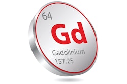
MRI effectively produces soft tissue images; it is also used to characterize active tumors with gadolinium-enhanced T1-weighted MR images. But gadolinium has been linked to health concerns -- namely, deposition of the agent in the brain and body -- and acquiring these contrast-aided sequences increases overall exam times and thus healthcare costs. That's why "deep learning (DL)-powered MR contrast synthesis emerges as a promising solution," Umapathy said.
To that end, the investigators used deep learning to synthesize T1-weighted contrast-enhanced MR images that incorporated tissue-specific information from unenhanced multi-contrast MR images (T1-weighted, T2-weighted, and FLAIR [fluid-attenuated inversion recovery] protocols). The MR contrast synthesis framework Umapathy and colleagues used is called Constrained Contrastive Learning-Synthesis (CCL-Synth). For the study, the team included 2021 Brain Tumor Segmentation Dataset information from 500 individuals; of these, 350 made up a training set, 80 made up a validation set, and 70 made up a test set.
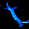
 Comparison of T1-CE images generated by the CCL-Synth model with ground truth and baseline results for three subjects. Yellow arrows indicate key regions where the CCL-Synth model achieves improved tumor conspicuity, enhancing visualization of tumor margins and internal structures compared to the baseline. Images and caption courtesy of Lavanya Umapathy, PhD, and the ISMRM.
The study's overall finding was that integrating tissue-specific MR contrast information in the synthesis of contrast-enhanced images improved image-quality evaluation metrics such as the structural similarity index measure (SSIM), the peak signal-to-noise ratio (PSNR), and learned perceptual image patch similarity (LPIPS), Umapathy said, noting that the approach "leverages multicontrast MR images to capture tissue-specific information through a self-supervised contrastive learning framework."
"Despite the technical challenges in synthesizing accurate and high-quality images, our deep learning approach demonstrated the feasibility of generating reliable T1-weighted contrast-enhanced images without the need for gadolinium injection," she concluded. "By leveraging prior information about underlying tissues through a self-supervised pre-training approach, our synthesis model achieves high perceptual quality and fidelity in synthesized images."