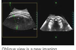
Contrast-enhanced ultrasound (CEUS) is more sensitive than contrast CT in detecting slight tumor blood flow in solid renal tumors, according to research published in the December issue of the Journal of Ultrasound in Medicine.
"In the evaluation of solid renal lesions, CEUS is less invasive than contrast CT and can show even slight tumor blood flow," wrote a team of Japanese researchers. "This imaging technique shows both tumor staining and vascular structures in detail and may be useful in patients in whom contrast CT or contrast MRI is contraindicated" (JUM, December 2005, Vol. 24: 12, pp. 1635-1640).
To evaluate the usefulness of CEUS in the diagnosis of solid renal tumors, a team of researchers from Wakayama University Hospital in Wakayama, Japan evaluated 29 patients with solid tumors detected on grayscale sonography between March 2002 and October 2004. All patients received further evaluation with arterial phase contrast CT and CEUS, and suspected malignant tumors were resected.
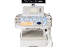
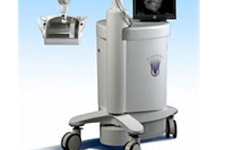
CEUS was performed using a Sonoline Elegra scanner (Siemens Medical Solutions, Malvern, PA), using the system's Sie Flow mode and a 2.5-g intravenous bolus of Levovist (Schering, Berlin, Germany). CT studies were given on an Aquilion multidetector-row CT scanner (Toshiba Medical Systems, Tokyo) at 120 kV (peak) and a revolution time of 0.5 seconds; patients received a 100 mL dose of iopamidol 300 (Bracco, Milan, Italy).
CEUS studies were reviewed by an internist, while contrast CT studies were reviewed by a radiologist. Findings were compared with the histopathological diagnosis.
Histopathological examination of the resected lesions found malignant tumors in 26 patients, including 18 cases of clear cell carcinoma, six cases of papillary renal cell carcinoma, one case of collecting duct carcinoma, and one case of infiltrative urothelial carcinoma. Three patients had benign tumors, including two cases of oncocytoma and one case of angiomyolipoma.
Contrast CT failed to show tumor blood flow in five of 29 patients, while CEUS showed tumor blood flow in all patients, according to the researchers. CEUS yielded a positive predictive value of 100%, compared with 82.8% for contrast CT.
In clear cell carcinoma cases, hypervascularity was shown on contrast CT in 16 of 18 patients and on CEUS in 17 of 18 patients, according to the authors. Based on hypervascularity, CEUS produced a diagnostic sensitivity of 88.9% and a specificity of 45.5% for clear cell carcinoma. Contrast CT yielded sensitivity of 88.9% and a specificity of 72.7%
"Contrast-enhanced ultrasonography therefore had better sensitivity but worse specificity than contrast CT," the authors wrote.
For papillary cell carcinomas, contrast CT revealed avascular lesions in four of six patients, with the radiologist diagnosing hemorrhagic simple cysts in these cases. However, CEUS showed blood flow in these lesions, indicating hypovascular renal tumors, according to the study team.
"CEUS is more useful in diagnosing hypovascular renal tumors than in diagnosing hypervascular renal tumors such as clear cell carcinoma," the authors wrote.
By Erik L. Ridley
AuntMinnie.com staff writer
November 24, 2005
Related Reading
RF ablation promising against small renal tumors, August 17, 2005
MRI-guided cryoablation promising against renal tumors, August 5, 2005
Renal mass with nonrenal malignancy may not need biopsy, December 2, 2004
Small size no predictor of benign renal masses, December 2, 2004
US, MRI challenge CT for classifying renal cell carcinoma, June 17, 2003
Copyright © 2005 AuntMinnie.com