Last year's international outbreak of severe acute respiratory syndrome (SARS) may feel like a distant memory, but some of the most thoughtful analyses of this new disease are just now emerging in medical literature.
One lesson is clear from several new studies: When the next outbreak occurs, plain x-rays can play a critical role in managing the crisis, distinguishing SARS-infected patients from those who merely have similar symptoms, and highlighting those who require intensive treatment to prevent mortality.
Specifically, an abnormal chest radiograph with haziness or pneumonia in the context of an outbreak is a high-risk predictor of SARS, according to one study. Also, more extensive airspace disease on radiographs at presentation is an independent predictor of adverse outcome in patients with SARS, according to a second study.
While the spate of cases in Toronto demonstrated the potential for a global epidemic involving the highly infectious disease, the latest literature comes from Hong Kong, which suffered the world's largest outbreak beginning in March 2003.
The absence of SARS cases in recent months is not taken for granted by the developers of a new clinical prediction rule published in the Annals of Internal Medicine (September 7, 2004, Vol. 141:5, pp. 333-342).
"The resurgence of SARS remains a distinct possibility in the coming winter months, given its uncertain origins and possible animal reservoirs (in the form of wild animal markets that are common in Guangdong, China)," wrote the authors from the Hospital Authority SARS Collaborative Group.
The Hospital Authority is responsible for all 44 public hospitals in Hong Kong, including those that provided clinical care for all the SARS patients in last year's outbreak. The Collaborative Group developed its prediction rule using data from 2,649 consecutive patients seen at two Hong Kong triage clinics during that time.
Of the 1,274 patients evaluated at United Christian Hospital, 377 (29.6%) were ultimately diagnosed with SARS; among the 1375 patients seen at the Prince of Wales Hospital, 184 (13.4%) had SARS.
The researchers performed a statistical analysis to assign positive or negative SARS risk scores to relevant patient variables. They divided the resulting prediction rule into two steps for the clinical decision process: a step 1 scoring system for initial triage based on patient history and symptoms, and step 2 for further radiographic and laboratory-test evaluation of patients deemed at high risk of SARS based on step 1.
An abnormal radiograph with evident haziness or pneumonia turned out to carry the highest risk score in step 2, with a score of 8 for that variable alone. A score of 8 or greater was correlated with high risk, as that cutoff achieved 95% sensitivity for a final diagnosis of SARS.
The only other equal or higher risk variables were seen in step 1, in which fever was assigned a risk score of 15, and contact with a SARS-infected patient garnered a risk score of 8. A total score of 3 or greater at step 1 was correlated with high risk for SARS.
The investigators also developed a scheme for classifying high-risk cases after step 2, so that patients could be appropriately assigned to different types of isolation wards.
"This process of allocation may be an important consideration in a large outbreak in which the surge capacity of hospitals would not be able to provide individual isolation rooms for all patients suspected of having SARS," the authors wrote, noting that isolation was "a major issue in the 2003 Hong Kong epidemic."
Using x-rays to determine which patients are at highest risk among the many who might become ill in a SARS outbreak is also the theme of a new article posted online prior to print publication in Radiology (September 16, 2004).
Researchers from the Prince of Wales Hospital and The Chinese University of Hong Kong, including lead radiologist Dr. Jeffrey K.T. Wong, retrospectively reviewed the radiographs of 138 healthcare workers and patients who contracted SARS and were treated at the hospital during last year's crisis.
The 66 men and 72 women included 32 patients (23.2%) whose infection progressed to acute respiratory failure requiring ICU admission, 19 (13.8%) who required invasive mechanical respiration support, and 8 (5.8%) who died from severe respiratory failure. All 138 underwent frontal chest radiography at initial presentation and during treatment using a portable CR unit (Mobilett Plus, Siemens, Erlangen, Germany).
A total of 2,045 chest x-rays (an average of 14.8 per patient) were obtained using a standardized technique: 75 kVp, 4 mAs, and a 180-cm film-focus distance for the posteroanterior views (taken when patients were able to stand up), and 70 kVp, 4 mAs, and a 100-cm film-focus distance for the anteroposterior views.
The article reports the findings of three radiologists who performed a consensus retrospective review of the x-rays while blinded to the patients' clinical progress.
"In view of the substantial number of patients with SARS who required ICU care, it would be desirable to have some parameters (clinical, laboratory, or radiologic) early in the disease that would help predict the clinical outcome," authors stated. "The extent of pneumonia at presentation radiologically appears to correlate with an adverse clinical outcome of SARS."
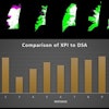
"There appears to be a strong correlation between the extent of radiographic abnormality and the degree of respiratory failure, as indicated by the Kaplan-Meier curve data, which show that even when only a small percentage (10%) of the lung was consolidated, approximately 50% of the patients required supplementary oxygen," the authors wrote.
The pattern of disease progression seen on radiographs was also highly correlated with the clinical outcome, according to the researchers.
"Patients with the type 1 progression pattern (initial radiographic progression followed by improvement) on serial chest radiographs seemed to have a more favorable outcome, whereas all seven patients with the type 4 pattern (progressive deterioration) had an adverse clinical outcome," the authors wrote. "Six of them died, and the seventh patient was still critically ill in the ICU at the time of this writing."
By Tracie L. Thompson
AuntMinnie.com staff writer
October 1, 2004
Related Reading
Mobile CT proves invaluable in SARS outbreak, January 22, 2004
Thin-slice CT reveals severe lung abnormalities in SARS patients, July 7, 2003
SARS presents distinct radiological patterns, May 6, 2003
Toronto SARS outbreak throws routine imaging into turmoil, April 18, 2003
Hong Kong hospital posts SARS protocol, March 25, 2003
Copyright © 2004 AuntMinnie.com