Postoperative ultrasound can play a useful role in detecting occult malignancies after breast cancer surgery, according to an article in the Journal of Ultrasound in Medicine.
"Surveillance ultrasonography, as an adjuvant to mammography, is helpful for the detection of an occult malignancy, clinically and mammographically, during the postoperative follow-up," according to a study team from Samsung Medical Center in Seoul, South Korea.
To investigate the efficacy of ultrasound in this application, the researchers performed 3,329 bilateral whole-breast ultrasound examinations in 1,968 asymptomatic patients after breast cancer surgery between January 2001 and February 2004 (JUM, May 2005, Vol. 24:5, pp. 643-649).
Ultrasound was performed at intervals of six months or more, combined or alternated with mammography. The studies were done using a Logiq 700 scanner (GE Healthcare, Chalfont St. Giles, U.K.) or a HDI 5000 system (Philips Medical Systems, Andover, MA) with a 5- to 12-MHz linear-array transducer by one of three board-certified radiologists or one of the institution's senior residents. Patients received scanning in their breast areas, chest walls, and axillae of both sides, as well as parasternal and supraclavicular regions of the treated side, according to the researchers.
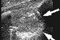
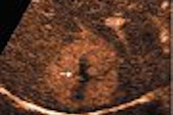
Aspiration or an ultrasound-guided core-needle biopsy was performed immediately or up to one week later on all positive examination results requiring pathologic confirmation. Of the 1,968 patients, 57 (2.9%) had a positive examination, yielding 24 malignant lesions (42%) and 33 benign lesions (58%).
None of the malignant lesions showed definite abnormalities on the concurrent or previous mammograms at the time of the ultrasonographic examination.
Ten false-negative cases were also found, four of which were identified by mammographic calcifications. Three more were found by enlarged internal mammary (two) and supraclavicular (one) lymph nodes on the chest CT. Two other cases were palpated by the clinician or the patient, and the remaining case was found by the hot uptake on a PET scan, the study team stated.
Overall, postoperative screening ultrasound yielded a sensitivity of 70.6% and a specificity of 98.3%.
"In our experience, ultrasonography is a useful surveillance method that can overcome the limitations and discomfort associated with a mammogram in those patients treated for breast cancer," the authors wrote. "Magnetic resonance imaging is known to be a highly sensitive modality for the recurrence of breast cancer; however, ultrasonography is more beneficial because of its high specificity, widespread availability, easily accessible biopsy procedures, comfort, and lower cost."
By Erik L. Ridley
AuntMinnie.com staff writer
May 4, 2005
Related Reading
Hormonal variations can affect breast vascularity, US reveals, January 13, 2005
Tumor type, breast profile determine value of mammo, US, and MR, January 6, 2005
Breast US readers need more practice with postop changes, December 22, 2004
Breast imagers fare better with hands-on approach to screening ultrasound, December 17, 2004
ACRIN updates multiple breast cancer trials, April 15, 2004
Copyright © 2005 AuntMinnie.com