Ultrasound's primary role in breast imaging is that of differentiating cystic from solid lesions detected by mammography. The modality can also be used to confirm a suspected or palpable mass, as an aid to treatment planning, and for needle guidance in interventional procedures. But it probably shouldn't be used for general breast screening.
"Ultrasound should be a directed study, so try not to screen for pathology," suggested Dr. Debra Ikeda in a presentation at the 2001 Royal Australian and New Zealand College of Radiology meeting in Melbourne, Australia.
Ikeda, an associate professor of radiology at Stanford University in Stanford, CA, offered some tips and techniques for ultrasound of the breast.
Compression
Compressing the breast tissue while scanning can reduce the number of false positives. Ikeda recommended compressing the breast tissue with the transducer in order to align the Cooper’s ligaments parallel to the transducer face. Cooper’s ligaments are fibrous structures that support the breast tissue and cause an acoustic shadow similar to that caused by malignant lesions.
Ultrasound images should be carefully labeled right or left, and radial sections should be identified using the clock face. The distance between any lesions and the nipple should also be measured and documented. Whether the scan plane is radial or antiradial also should be indicated. All of this information is necessary for generating an accurate report, and is helpful if the patient returns for an ultrasound-guided procedure, Ikeda said.
Masses, lesions, and cysts
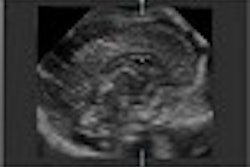
"Malignant lesions will be taller than they are wide," Ikeda said. They are also hypoechoic, with angulated margins, cause an acoustic shadow, show duct extension, and have irregular borders.
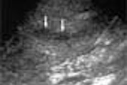
Cysts without smooth borders may represent irregular mucinous carcinomas. Regions of increased echogenicity often surround tumors, and this represents peritumoral reaction. Ikeda recommended scanning from many directions and to "smash down the tissue" to reduce any false shadowing. In her department, a cotton swab or large paper clip under the transducer are used to mark the position of the suspected lesion. This will cast an acoustic shadow in the ultrasound image and enable a better assessment of the area of interest.
For palpable masses, ultrasound can be used to distinguish malignant from benign.
"Listen to the patient and scan over the area they are concerned about for reassurance," Ikeda advised.
The only remaining problem, then, is with isoechoic lesion. These lesions are inconspicuous because they have the same echogenicity as the surrounding breast tissue. A gel standoff may be necessary in very superficial lesions to achieve improved image quality in the near field and demonstrate any changes in skin contour, she said.
Difficulties arise after biopsy, lumpectomy, and chemotherapy in the region of cavities and scar tissue. Ultrasound will show classic distortion of the skin and fat relating to the cavity, as well as architectural distortion and thickened skin. She recommended that typically benign lesions should undergo follow-up, not biopsy.
Pregnant patients
There is a greater risk of breast cancer during pregnancy, and during this time, the breasts are bigger and lumpier. Breast lesions may be masked on mammography. Ikeda warned that delayed detection worsens the prognosis.
In these patients, ultrasound should be performed first. The noncompressible (abnormal) breast will look smaller compared with the contralateral breast due to a lack of breast tissue spread, and this is an important, but often overlooked, indicator of pathology, she indicated.
With the advent of MRI, ultrasound is being used to confirm MR findings. "Directed ultrasound showed 9 out of 10 lesions shown on MR that were not demonstrated on mammography," she said. MR-guided biopsy, using an MR-compatible needle, is an alternative for lesions that are isoechoic on ultrasound, she said.
By Leanne McKnoultyAuntMinnie.com contributing writer
January 22, 2002
Leanne McKnoulty is an accredited medical sonographer currently lecturing in ultrasound at Monash University in Melbourne, Australia. She also is an editor at Ultrasound Review.
Related Reading
Ultrasound aids mammography in breast cancer diagnosis, December 3, 2001
New ultrasonic approach focuses on breast imaging, November 27, 2001
Ultrasound marks metastatic lymph nodes for breast cancer staging, October 17, 2001
Copyright © 2002 AuntMinnie.com