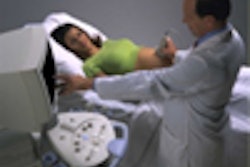
Using the fontanelle and sutures as an acoustic window, 3-D ultrasound offers physicians a detailed look at the fetal brain. Dr. Ilan Timor-Tritsch discussed the value of transvaginal ultrasound for this technique at the 2001 International Society of Ultrasound in Obstetrics and Gynecology conference in Melbourne, Australia.
In cephalic presentations, the head is manipulated into the desired position by pressing on the maternal abdomen with the nonscanning hand, said Timor-Tritsch, who is the director of ultrasound at the New York University School of Medicine in New York City.
"The 3-D images we more commonly see of the fetal face use a surface-rendered technique that displays the outline of anatomy such as the skin, tongue, and eyes. Improved prenatal characterization of facial clefts, particularly of the palate, have been demonstrated using surface rendering. Sophisticated 3-D diagnosis of fetal brain abnormalities is achieved using multiplanar reconstruction," he said. "This enables visualization of three ultrasound planes simultaneously."
The Voluson 530D 3-D system (Medison America, Cypress, CA) that Timor-Tritsch uses offers real-time 3-D images and can automatically acquire three orthogonal planes of ultrasound data. Using multiplane exploration, it is possible to travel inside the cube and achieve precise creation of any chosen section planes, including the axial plane that is perpendicular to the ultrasound beam.
 |
"The 3-D volume could be examined using the classical parallel sections in all three orthogonal planes," he said. These planes are used in both CT and ultrasound and are more easily interpreted by other clinicians, such as neurologists and neurosurgeons.
"They were able to provide a more focused and precise postnatal management based upon the objective evaluation of the pathology," Timor-Tritsch reported.
Using the 3-D volume as a guide, the user navigates through the stored information within the "active box" by adjusting the marker dots at the side of the image that indicate position.
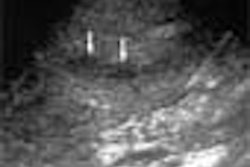
"This dot is generated by the intersection of three planes, is freely movable by the user, and marks the same spot within the volume," he said. It is possible to simultaneously view a structure such as a choroid plexus cyst in the sagittal, coronal, and axial planes. It is then possible to move up, down, or across the volume, and the three ultrasound planes automatically adjust to the new area of interest."
In addition, there are a number of neurological abnormalities that are more easily and confidently diagnosed using transfontanelle scanning. In his presentation, he demonstrated a number of such cases, including agenesis of the corpus callosum, hydrocephaly, alobar holoprosencephaly, and some hard-to-define cases such as arachnoid cysts. In the case of agenesis of the corpus callosum, it was evident on the midcoronal plane that the corpus callosum was missing and the lateral ventricles dilated.
"We were able to trace in all three planes the location of the upwardly displaced third ventricle and assure ourselves that the corpus callosum was indeed missing," he reported.
Using 3-D for diagnosing hydrocephaly enabled a more comprehensive understanding of the extent of ventricular enlargement, and neurosurgeons found it easier to picture, Timor-Tritsch added.
In another case of partial agenesis of the corpus callosum, a power angiography mode was used to position the marker dot, which enabled the anteriorcerebral artery and its branch, the pericallosal artery, to be followed. These pericallosal arteries act as an important indicator that the corpus callosum is absent, he said.
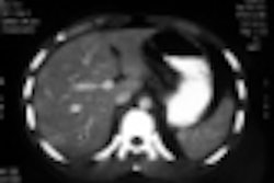
The third mode of 3-D operation is known as transparent mode, which strips off soft tissue and highlights bony structures such as the spinal column. Timor-Tritsch recommended using the 12th rib as an anatomical reference, and said he has used this mode for easy detection of spinal abnormalities, including hemivertebrae. Conversely, it is possible to extract only the anechoic structures and recreate cystic structures such as ventricles and blood vessels, he explained.
"3-D is the icing on the cake when it comes to defining difficult pathology," he concluded. "In the future, we’ll use 3-D for all neurosonograms. Now that we have it, we definitely can’t do without it."
By Leanne McKnoultyAuntMinnie.com contributing writer
December 18, 2001
Leanne McKnoulty is an accredited medical sonographer currently lecturing in ultrasound at Monash University in Melbourne, Australia. She also is an editor at Ultrasound Review.
Related Reading
Fetal nasal bone examination may improve Down's screening, November 16, 2001
Multinational study makes case for first-trimester sonography, June 12, 2001
Copyright © 2001 AuntMinnie.com