Contrast-enhanced ultrasound (CEUS) can offer significant value in differentiating breast masses initially categorized as ultrasound BI-RADS category 4 by improving specificity, according to Chinese researchers who presented the study at this month's RSNA meeting in Chicago.
A Chinese research team from Huashan Hospital in Shanghai found that CEUS led to a significant improvement in specificity and accuracy over conventional ultrasound in lesions initially categorized as ultrasound BI-RADS 4.
"CEUS has potential in [leading to a] better therapeutic plan with [fewer] unnecessary interventional diagnosis or treatment," said presenter Dr. Ping Xu.
Since ultrasound BI-RADS 4 lesions cover a wide range of malignant possibilities (3%-94%), the research team sought to investigate the use of real-time grayscale CEUS to recategorize the lesions.
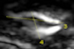
In their study, 31 patients with 31 breast lesions received conventional ultrasound on an Acuson Sequoia 512 scanner (Siemens Healthcare, Erlangen, Germany), and were categorized as BI-RADS category 4. CEUS was also performed following intravenous bolus injection of the SonoVue contrast agent (Bracco Imaging, Milan, Italy).
Blinded to the conventional sonographic findings, two other investigators observed and analyzed tumor size, enhancement pattern, time-intensity curve, and contrast parameters, according to Xu. Based on literature review, contrast enhancement patterns of malignant breast lesions were defined as:
- Poorly-defined margin
- Penetrating or tortuous surrounding vessels
- Heterogeneous distribution of the contrast media
- Apparently larger size of the lesion at CEUS
- An obvious increase in peak intensity (PI)
The two investigators categorized the lesions as BI-RADS 5 (highly suggestive of malignancy) if the first three patterns were present and as BI-RADS 4 (suspicious abnormality) if any of the five patterns were seen. When none of the conditions were present, the investigators classified the lesions as BI-RADS 3 (probably benign).
Based on the CEUS data, 11 of the 31 lesions were categorized as BI-RADS 5, 16 as BI-RADS 4, and four as BI-RADS 3. All 31 masses were excised; 21 lesions were malignant and 10 were benign.
CEUS performance in BI-RADS 4 lesions
|
In other findings, CEUS accurately classified 10 of 11 (90.9%) of the lesions categorized as BI-RADS 5 lesions, 12 of 16 (75%) of lesions categorized by BI-RADS 4, and all four lesions (100%) categorized as BI-RADS 3.
"We found that CEUS is useful for the differentiation of BI-RADS 4 lesions," she said.
By Erik L. Ridley
AuntMinnie.com staff writer
December 23, 2009
Related Reading
Contrast ultrasound may spot early signs of atherosclerosis, October 20, 2009
Contrast US beats MRI, CT for liver lesions, August 17, 2009
Interest grows in noncardiac ultrasound contrast applications, May 21, 2009
CEUS of focal liver lesions in the noncirrhotic liver, May 19, 2009
Contrast ultrasound beats CT, PET in Hodgkin's lymphoma staging, May 6, 2009
Copyright © 2009 AuntMinnie.com