CHICAGO - Elastography has the potential to reduce unnecessary biopsies, according to a study presented at the RSNA meeting on Sunday.
Researchers from Elizabeth Wende Breast Care in Rochester, NY, found a correlation of 98% between elastography, standard ultrasound, clinical mammography, and biopsy for cancerous lesions. For benign lesions, although the percentage was a bit lower at 78%, elastography could still help clinicians decide whether biopsy is needed.
"We see a lot of young women in our center who've felt a lump and come in to have it checked out," said the study's lead author, Dr. Stamatia Destounis. "Typically, these are fibroadenomas, and if we trust elastography, we don't need to perform biopsies for lesions like that. We can avoid unnecessary testing."
Elastography improves ultrasound's specificity by using conventional ultrasound to measure the compressibility and mechanical proportions of a lesion. The idea is that because cancer tissue tends to be stiffer than healthy tissue, if a lesion is compressible it's less likely to be malignant. The technique's measurements for cancerous lesions tend to be smaller than ultrasound's, and its measurements for benign lesions tend to be larger than ultrasound's, Destounis said.
The researchers used a Sonoline Antares in the beginning of the study, before upgrading to an Acuson S2000 unit (both from Siemens Healthcare, Erlangen, Germany). Between 2008 and 2009, an elastography exam was given at the same time as a hand-held ultrasound exam to 193 patients with 198 lesions. All solid lesions had some form of needle aspiration. Destounis and colleagues compared standard B-mode ultrasound, elastography, cine loops, and color mapping, and collected data for the following:
- B-mode lesion size
- Elastography lesion size
- Percentage likelihood of malignancy, based on 2D ultrasound versus elastography
- Lesion ratios (side to side, top to bottom)
The team performed 140 biopsies. Out of those 140 biopsies, 59 cancers were found, according to Destounis.
"We were able to show concordance between B-mode, elastography, and needle biopsy in 98% of those cancer cases," she said.
 |
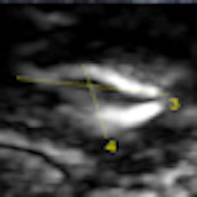
| 46-year-old patient presents for evaluation of left breast nodule after being recalled from screening mammography. Above, mammogram: Nodule present within the left central aspect. Below, B-mode and elastography: Ultrasound shows a cyst present corresponding to area noted on mammogram. Elastography image demonstrates the "bull's-eye" appearance of cysts on elastography. A cyst aspiration was performed and the cyst resolved. Images courtesy of Dr. Stamatia Destounis. |
 |
No consensus yet
Despite these positive results, not everyone agrees that elastography is trustworthy enough at this point in its development. In another Sunday presentation, Korean researchers described a study they had conducted with 284 women with 342 sonographically visible breast lesions scheduled for biopsy between December 2007 and December 2008. They also used Siemens' Sonoline Antares, and compared elastography's ability to characterize lesions with that of conventional ultrasound.
Dr. Jin Hwa Lee from Dong-A University in Busan, South Korea, and colleagues found that elastography's specificity was 83.3%, compared to 28.4% for B-mode ultrasound. But its sensitivity was far lower at 77.4%, compared to 98.8% sensitivity for B-mode, leading the researchers to conclude that the technology's advantages may not overcome the risk of missed cancer.
The differing results are probably due to elastography's ongoing refinement, Destounis said. She estimates that the technique will go mainstream -- as a built-in addition to future ultrasound units -- over the next four to five years.
"Elastography is definitely not the end-all-be-all," she said. "It's another tool, and you don't decide not to biopsy something just because of elastography results -- you have to use all the clinical information that you have. The technology is progressing, with multiple vendors trying to perfect it. It's a developing technique."
By Kate Madden Yee
AuntMinnie.com staff writer
November 30, 2009
Related Reading
Elastography shines in assessing breast lesions, April 29, 2009
Adding sonography to yearly mammograms helps find metachronous cancers, February 25, 2008
Breast sonoelastography aids in evaluating breast lesions, January 28, 2008
Elasticity imaging finds breast tumors based on shape, volume, December 14, 2007
Copyright © 2009 AuntMinnie.com