ATLANTIC CITY - A subharmonic contrast-specific imaging technique can offer specificity advantages over mammography for breast cancer diagnosis, according to a pilot study from Thomas Jefferson University (TJU) in Philadelphia.
After completing initial research on subharmonic imaging (SHI) on animals, TJU researchers performed a human study to compare grayscale SHI to conventional grayscale and power Doppler imaging (PDI), with and without contrast, as well as mammography for breast cancer diagnosis. Flemming Forsberg, Ph.D., presented the institution's research during a talk at this week's Leading Edge in Diagnostic Ultrasound conference.
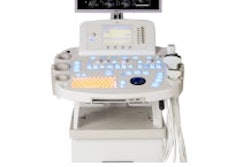
The study included 14 women with 16 breast lesions who were scheduled for core or surgical biopsy. The women received a baseline grayscale ultrasound study, a pre- and postcontrast power Doppler study, and grayscale SHI using a modified Logiq 9 scanner and 7L probe (GE Healthcare, Chalfont St. Giles, U.K.).
Six women received Optison (GE Healthcare) prior to the agent's recall in late 2005, while eight received Definity (Bristol-Myers Squibb Medical Imaging, North Billerica, MA). Ultrasound exams were read by one reader, with diagnoses assessed on a six-point scale (no lesion to definitely malignant).
Ultrasound diagnostic criteria included size, shape, and orientation; echogenecity; degree of enhancement; tumor margin definition; through transmission characteristics; degree and pattern of vascularity; blood vessel characteristics; and vessel anastamoses. Digital clips were transferred to a PC, and SHI time-intensity curves were calculated within each lesion, according to Forsberg.
The researchers calculated sensitivity, specificity, and accuracy rates against pathology results using McNemar's test. Of the 16 lesions, four were malignant and 12 were benign.
Mammography was performed on 11 patients (13 lesions all with BI-RADS 3 and 4 findings), with MRI, ultrasound, and palpation each for one subject. Pathology results were available for 13 patients, with the last subject diagnosed via follow-up, Forsberg said.
For breast cancer diagnosis, the sensitivity, specificity, and accuracy rates for each study type are shown below.
|
The differences in accurate rates were only statistically significant between SHI and mammography (p = 0.07), according to Forsberg.
In other findings, no diagnostic criteria correlated with pathology, he said. Also, the degree of vascularity observed with PDI and with contrast PDI both correlated with SHI, but not with each other, he said. Also, SHI signals were 100% likely to produce good or excellent enhancement, compared with 44% for PDI contrast signals (p = 0.004).
"The specificity of all the ultrasound modes was significantly higher than mammography and the accuracy of SHI was marginally higher than mammography," Forsberg said.
The area under the ROC curve for breast cancer diagnosis was highest with SHI (0.78, p > 0.1). All ultrasound modalities combined produced the highest area under the ROC curve (0.87, p > 0.1).
Forsberg cautioned that the pilot study was based on a very limited dataset. "These conclusions are therefore only preliminary," he said.
The institution is seeking funding, however, from the National Institutes of Health (NIH) for further research on a larger patient population, Forsberg said.
By Erik L. Ridley
AuntMinnie.com staff writer
May 26, 2006
Related Reading
Breast surgeon expresses reservations about screening US, April 13, 2006
Auto breast US handily spots lesions in early study, January 13, 2006
Lesion size, diameter, and location affect breast US interpretation, November 28, 2005
MRI best for familial breast cancer detection, December 16, 2005
Copyright © 2006 AuntMinnie.com


