Sonography with microconvex probes is suitable as a first-line diagnostic method for diagnosing meniscal tears, according to research published in the May issue of the Journal of Ultrasound in Medicine.
"We recommend high-resolution microconvex probes, which better fit the anatomic concavity of the popliteal fossa, as efficient investigation tools," wrote researchers from Shaheed Beheshti Medical Science University in Tehran, Iran (JUM, May 2006, Vol. 25:5, pp. 593-597).
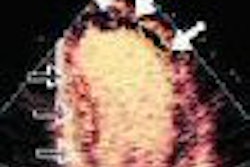
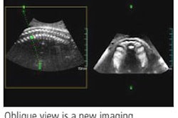
The study team employed a 6.5-MHz microconvex probe to prospectively evaluate 408 knee joints with knee pain and a clinical indication for arthroscopy. The sonographic study started from the midline, with the probe smoothly moved toward the medial and lateral aspects of the popliteal fossa, according to the authors.
Both sagittal and transverse images were obtained. The hypoechoic area in the center of the echogenic meniscus was considered an interstitial tear and degeneration; hypoechoic lines extending to the superior or inferior margin of the meniscus were considered meniscal tears. The posterior horns of both menisci were examined, according to the authors.
All exams were performed by a trained musculoskeletal radiologist. Of the 406 patients, 100 had positive sonography findings and underwent an arthroscopic examination. The findings were then compared.
Sixty medial meniscal tears and 47 lateral meniscal tears were demonstrated on sonography, compared with 58 medial meniscal tears and 47 lateral meniscal tears on arthroscopy.
Sonography produced sensitivity of 100% and specificity of 95% in detecting meniscal tears, according to the researchers. For the medial meniscus, sonography yielded a positive predictive value of 95% and a negative predictive value of 100%. The modality produced a positive predictive value of 93% and a negative predictive value for lateral meniscal tears.
As a supplement to clinical findings, sonographic examination of the meniscus is an easily available, noninvasive, and time- and cost-efficient imaging technique that can be used instead of MRI to optimize preoperative diagnosis and to determine indications for arthroscopy, the authors concluded.
"The use of microconvex probes provides direct contact with the posterior horns of menisci and easier application regarding their shape and size," the authors wrote. "Careful consideration of the technical requirements and systemic performance of the dynamic examination should lead to further improvement of examination results, even in bucket handle tears, and to growing clinical effectiveness in the future."
By Erik L. Ridley
AuntMinnie.com staff writer
May 10, 2006
Related Reading
Meniscus damage contributes to cartilage loss in knee osteoarthritis, March 10, 2006
Four MRI signs diagnose radial meniscal tears preoperatively, February 23, 2005
Fast spin-echo MRI unsuitable for meniscal tears, July 6, 2005
Studies critique radiologists’ reports in chest x-ray, knee MRI, April 19, 2005
Meniscal MRI studies highlight axial advantages, radial tear rates, August 28, 2004
Copyright © 2006 AuntMinnie.com