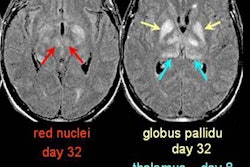
Most cases of pediatric non-Hodgkin’s lymphoma (NHL) fall into three histologic subtypes: undifferentiated, lymphoblastic, and large cell, with each subtype having typical clinical and imaging features. CT is the primary modality used to evaluate these tumors, along with skeletal scintigraphy and ultrasound. MRI is used mainly for cancer staging and when there is suspicion of central nervous system or bone involvement.
Researchers at The Hospital for Sick Children in Toronto, however, are using whole-body MRI to stage pediatric NHL. Their findings thus far have been encouraging, and they believe MRI may one day replace standard radiographic procedures.
"Based on our initial experience, whole-body MRI in pediatric patients can detect lymphomatous involvement not only of bone marrow, but also of lymph nodes and parenchymal organs," said Dr. Christian Kellenberger.
Kellenberger presented the results of a preliminary study on whole-body MRI for NHL staging at the 2002 RSNA conference in Chicago. In October 2001, eight pediatric patients at his institution with confirmed diagnosis of lymphoma underwent whole-body MRI to assess bone marrow involvement. Six of the patients had NHL and two had Hodgkin’s disease.
Whole-body MRI was performed as part of the initial staging in five patients and as follow-up in the remaining three. All the MR studies were conducted on a 1.5-tesla scanner using body coils. The entire body was covered by multiple coronal fast-spin echo inversion recovery (FSEIR) with a field of view of 300-480, a 320-512 x 160-256 matrix, slice thickness of 6-12 mm, a TR of 4000-4950, TE of 22-42, TI of 150, a NEX of 0.5-1, and an echo-train length of 4-10. Two patients were studied under general anesthesia and six were studied without requiring sedation.
"The MR images were assessed for any abnormality such as increased signal within bone marrow sub-tissue, any altered signal within parenchymal organs, or enlarged lymph nodes," Kellenberger said.
Foci of increased signal on FSEIR coronal images within marrow spaces (allowing for expected age-related changes), cortical bone, and parenchymal organs, as well as enlarged lymph nodes, were considered abnormal and highly suggestive of tumor, noted Kellenberger. Three observers classified each observed lesion as involved by lymphoma or as an incidental finding.
"A site was considered involved based on all available imaging and clinical information and histology," Kellenberger said.
MRI and standard imaging procedures were then compared for the detection of the sites considered involved by lymphoma. The standard staging procedures conducted at the institution included CT, skeletal scintigraphy, and gallium scintigraphy.
"When compared with standard staging imaging procedures, whole-body MRI findings correlated well with CT, skeletal scintigraphy, gallium scans, and bone marrow aspiration," Kellenberger said.
A total of 48 regions suspicious for tumor were seen on CT, 142 on MRI, 32 on gallium scintigraphy, and 12 on bone scan. The suspected diagnosis of non-Hodgkin’s lymphoma was confirmed in 6 of the 8 patients. Of the two children with Hodgkin’s disease, whole-body MRI findings also correlated well with standard imaging.
"Compared with standard imaging, whole-body MRI was more sensitive in detecting bone marrow lesions during initial staging, but was less specific for involvement of lymph nodes or of bone marrow in treated patients. This is likely due to use of only coronal sequences," Kellenberger said.
Although the preliminary results are encouraging and suggest that whole-body MRI may replace standard radiographic procedures for the staging and follow-up of NHL in children, the team is seeking to conduct further studies to determine accuracy and develop additional protocols.
By Jonathan S. BatchelorAuntMinnie.com staff writer
January 2, 2003
Related Reading
Child neurologists set guidelines for tests of global developmental delay, October 29, 2002
Pediatric sedation risk lowered when guided risk assessment performed, February 6, 2002
Melatonin puts kids to sleep, makes MRI a dream, December 10, 2001
Contrast agents herald new progress in MR lymphography, August 9, 2002
Contrast MRI improves cancerous lymph node detection, misses some primary tumors, October 31, 2002
Tracking of pediatric sedation for MR imaging highlights concerns, July 11, 2000
Copyright © 2003 AuntMinnie.com