Whole-body CT screening, the bête noir of consumer-driven healthcare, managed to nail potentially serious pathologies in more than a third of its asymptomatic subjects in the largest study of its kind to date.
Commensurate with today's ultracautious approach to the technique, the researchers strongly cautioned that more research is needed to determine the importance of the findings and assess the effects of whole-body screening on patient care. At any rate, the compendium of results is interesting inasmuch as it paints one of the most detailed pictures to date of what goes wrong with a population as it ages.
While imaging center operators have marketed whole-body CT directly to patients as an overall screening test, important questions have yet to be addressed, according to Dr. Claudia Furtado, Dr. Diego Aguirre, Dr. Claude Sirlin, and colleagues from the University of California, San Diego (UCSD).
"Some investigators have stated that whole-body CT screening is cost-effective on the basis of sparse data and controversial assumptions; other investigators have cited anecdotal experience to illustrate the potential pitfalls of CT screening, particularly the high number of false-positive test results and the consequences of them," the group wrote in the November issue of Radiology. "To our knowledge, no cost-effectiveness analysis or large-scale randomized trials have been performed for evaluation of whole-body CT screening" (Radiology, November 2005, Vol. 237:2, pp. 385-394).
The frequency of findings needs to be addressed before large-scale studies can be performed, the investigators stated. They retrospectively reviewed CT studies of 1,192 consecutive asymptomatic patients (774 men and 418 women, mean age 54) who underwent whole-body CT screening at a standalone for-profit imaging center in 2000. The UCSD researchers noted that there was no financial relationship between the imaging center and the academic researchers who compiled the results.
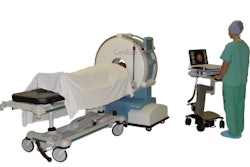
An electron beam scanner (C-150 XP, GE Healthcare, Chalfont St. Giles, U.K.) was used to acquire all data, typically 6-mm-thick sections at 200 mAs and 130 kVp. Standardized coronary calcium scoring was performed from the data, with the results interpreted by a cardiologist. Initial interpretation of the scans was performed remotely on a grayscale monitor and 3D reformation functionality (InSight Diagnostic Imaging Workstation, Neo Imagery Technologies, City of Industry, CA) by two board-certified and experienced radiologists who were not part of the research team, the authors wrote.
Demographic, diagnostic, and medical history information were also recorded at the time of imaging, while disease duration, stage, and severity were not consistently reported. Findings were categorized according to their anatomic location and included impressions and recommendations, if any, for follow-up. Statistical analysis included means and standard deviations reported for quantitative measures, and percentages reported for categorical data.
Findings in vast majority
According to the results, 1,030 patients (86%) had at least one abnormal finding described in the radiology report. In all there were 3,361 findings, a mean of 2.8 per patient. The 162/1,192 (14%) patients with no findings tended to be younger (mean age 43 years ± 9, p < 0.01) than the 1,030 who were positive for at least one finding (mean age 56 years, ± 10), and the ratio of findings per patient increased in a stepwise fashion with advancing age (p < 0.001). Yet the prevalence of findings in men (668 [86%] of 774) versus women was otherwise similar.
As for anatomic location, the distribution was similar for each age group; abdominal findings were the most common and thoracic findings were the least common. There were 779 noncardiac findings, and 42% (505/1,192) had at least one noncardiac finding in the thorax.
In all there were 461 lung findings representing 59% of all thoracic findings. The most common among these were parenchymal scars (153/461 33%), indeterminate lung nodules (115/461, 25%), granulomas (71/461), and emphysema (42/461, 9%). One patient had a 4-mm mass.
The 271 mediastinal findings represented 35% of the thoracic findings; among these the most common were vascular abnormalities (n = 117), wall calcifications (n = 76), thoracic aortic enlargement (n = 26), and enlarged pulmonary arteries (n = 15). In addition there were 47 chest wall findings, 91% of which were breast findings.
A third of the findings were in the spine (1,065/3,3,361, 32%), followed by abdominal blood vessels (561/3,361, 17%), lungs (461/3,361, 14%), kidneys (353/3,361, 11%), and liver (183/3,361, 5%).
Among 1,065 findings of the spine, 583 (55%) were degenerative changes, 416 (39%) were abnormally low bone density, and there were 14 miscellaneous benign lesions, commonly pars defects. Three indeterminate sclerotic lesions were also reported, including an exophytic lesion in the L5 transverse process and two focal lesions in the L2 and left ilium, respectively.
Follow-up recommendations in more than a third
In all 445/1192 (37%) patients received recommendations for follow-up, in a pattern that was similar for men and women, but increased with the subject's age (11% younger than 40 years and 56% at 70 years and older). Follow-up imaging was the most common recommendation (400/562, 69%, most of whom were referred for another CT exam) among the mostly self-referred patients.
"Although the study sample consisted mainly of middle-aged adults, there was a wide range (22-85 years) among subjects who underwent screening," the authors wrote. Four percent of the patients who self-referred reported a history of malignancy, and 8% were under age 40 "and those in this age group have a low pretest probability of disease, and therefore, are unlikely to benefit from screening."
Also, a higher number of findings per patient was found in men, but women had a significantly higher prevalence of follow-up recommendations, both per patient and per finding, driven predominantly on the basis of findings in the breast and ovaries.
Sixty-nine percent of patients (826/1,192) had at least one finding in the abdomen or pelvis, including most common findings in the blood vessels (561/1,517, 37%), genitourinary tract (364/1,517, 24%), and hepatobiliary tract (230/1,517, 15%).
Calcification was by far the most common vascular abnormality (540/561, 96%), though more clinically important aneurysms counted for 2% (13/561), and there were 8/61 (1%) vascular ectasia. The 13 aneurysms included five aortic artery aneurysms (mean size 32 mm, range 15-48 mm), four iliac artery aneurysms (mean size 19 mm, range 13-23 mm), two renal artery aneurysms (size unreported), and two splenic artery aneurysms (13 and 23 mm).
Of 353 renal findings, 58/104 (56%) were single cysts and 46/104 (44%) were multiple cysts (mean size was 37 mm ± 27, range 10-100 mm).
The 230 hepatobiliary findings included 43 single hepatic cysts, 28 multiple cysts, and 31 single nonattenuating lesions. Dilated bile ducts were not measured and hepatic dimensions were unreported.
Male reproductive system findings included diffuse prostate enlargement (52/91, 57%), prostate calcification (20/91, 22%), and seminal vesicle or vas deferens calcification (9/91, 10%) but no suspicious testicular masses or abnormalities.
Female reproductive system findings included uterine fibroids (36/84, 32%) and ovarian cystic masses (27/84, 32%, mean diameter 35 mm ± 13).
Of 62 splenic findings, 87% (54/62) were calcifications, 11% (7/62) were cysts or low-attenuating lesions, and 2% (1/62) were mild splenomegaly, but no solid or indeterminate splenic masses were reported.
Gastrointestinal abnormalities included diverticuli (27/42, 64%), including 25 in the colon, one in the stomach, and one in the duodenum; wall thickening was seen in 8/42 (19%), among other findings.
Implications for the whole-body debate
On the minus side, "whole-body CT screening may conceivably alter patients' abilities to obtain medical, disability, or life insurance," the authors stated. "Other adverse effects associated with workup after positive results are obtained in asymptomatic patients include increased anxiety and potential complications from intravenous contrast material administration or invasive procedures ... that are performed for further characterization of encountered findings. These adverse consequences are particularly undesirable if the original screening results were false-positive, or if the natural course of disease is not improved with early detection." Also, false negatives could create a false sense of security, and radiation is another potentially adverse consequence of CT screening.
"Moreover, for screening to be cost-effective, two, among other, criteria must be met: (a) screening must help in the detection of preclinical disease and (b) detection of preclinical disease must translate into longer survival and/or higher quality of life."
Additional legal, social, and healthcare issues include contrast media reactions, the legal impact of false-negative exams, and complications that occur during follow-up exams.
"On the basis of our results, we believe that prior to advocating CT screening to the general public, further research is needed to optimize whole-body CT screening techniques, determine (its) diagnostic performance, standardize reporting, establish a system of patient stratification according to risk and demographic factors, fully address ethical and legal issues, create cost-benefit models, and, if still warranted, perform randomized clinical trials," the authors concluded.
"The results were predictable," stated Dr. Michael Brant-Zawadski, medical director of radiology at Hoag Memorial Hospital in Newport Beach, CA, but "they will raise the same old debate."
Still, the screening advocate noted in an e-mail to AuntMinnie.com that methods are in process that can help categorize and stratify the risks and benefits of various findings.
"With greater experience, the 37% recommendations for follow-up studies/procedures would likely decrease to 20% to 25% (e.g., see the latest ELCAP recommendations for pulmonary nodules and decreasing need for follow-up)," Brant-Zawadski wrote.
At any rate, he added, with more and more clinicians putting CT scanners in their offices, the screening CT is becoming an in-office physical. "It's out of radiology's hands," he stated.
By Eric Barnes
AuntMinnie.com staff writer
November 9, 2005
Related Reading
Do whole-body CT's costs outweigh the benefits? February 2, 2005
False-positive cancer screens common and costly, December 17, 2004
MDCT panel addresses CT screening issue, June 25, 2004
Is your preventive imaging program responsible? November 24, 2003
Mayo 'whole-body' screening keeps an eye on the brand, October 3, 2003
Copyright © 2005 AuntMinnie.com