Intracardiac echocardiography (ICE) does a respectable job of planning radiofrequency catheter ablation (RFCA) for atrial fibrillation, and offers some unique strengths, such as real-time imaging during the procedure. But multidetector-row CT held onto its gold-standard status in a new study from Europe, detecting additional veins and pulmonary venous (PV) branches compared to ICE, which also suffered from underestimation of PV ostial size.
"Radiofrequency catheter ablation is a potentially curative treatment modality for atrial fibrillation originating in the pulmonary veins," explained Dr. Monique Jongbloed, Dr. Jeroen Bax, and colleagues from Leiden University Medical Center in the Netherlands. "For proper targeting of radiofrequency lesions, accurate visualization and knowledge about PV anatomy is necessary" (Journal of the American College of Cardiology, February 1, 2005, Vol. 45:3, pp. 343-350).
ICE makes a strong candidate for RFCA planning because of its potential advantages, Jongbloed and colleagues wrote.
"Advantages of (ICE) include the possibility to obtain online information of left atrial anatomy and PV in relation to ablation catheters, potential to assess hemodynamic function, and the monitoring of acute complications, such as pericardial effusion and acute PV stenosis," they explained.
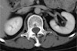
The aim of the study was to compare ICE to MDCT as the gold standard for providing anatomic information regarding PV anatomy in RFCA patients, and compare measurements of the ostia in 2D MDCT versus ICE. The study evaluated the PV and atrial insertion in 42 patients (35 men, 7 women, ages 49 ± 9 years) who were admitted for ablation of PV ostia.
CT was performed on either a four-slice (16 patients, 2-mm collimation) or a 16-slice (26 patients, 0.5-mm collimation) Aquilion scanner (Toshiba Medical Systems, Tokyo) following administration of 160 mL of nonionic contrast material (Xenetix 300, Guerbet, Roissy, France) at 4 mL/sec. Pitch was 1 mm/0.5 sec and 4 mm/05 sec for the four- and 16-slice scanners respectively, and tube voltage was 120 kVp at 250 mAs.
Three-dimensional reconstructions were viewed in 2D viewing modes on a Vitrea workstation (Vital Images, Plymouth, MN), and reconstructions were evaluated in the transversal, sagittal, and coronal planes, with 3D reconstructions used to evaluate PV anatomy.
ICE was performed using a 64-element monoplane phased-array transducer (AcuNav diagnostic ultrasound catheter) on a Sequoia ultrasound system (Siemens Medical Solutions, Malvern, PA). The catheter was inserted in the right atrium through the left femoral vein, PV anatomy was evaluated with ICE, and measurements of PV ostia were performed, the authors wrote.
According to the results, MDCT demonstrated common ostia of the left PV in 33 (79%) patients, compared to 31 (74%) of patients with ICE. Common ostia of the right PV specifically were seen in 13 (31%) patients with MDCT and seven (17%) patients with ICE. As for the subsequent RFCA, procedural success was achieved in 34/36 (94%) patients, with no major complications.
Ostial diameters measured in MDCT were similar to 2D ICE measurements in the anterior-posterior direction; however, MDCT measured significantly larger diameters in the superior-inferior direction compared to ICE. Yet the difference between all measurements performed with MDCT and ICE "was within two standard deviations from the average difference for most measurements, as was demonstrated with Bland-Altman analysis," the authors noted.
One problem with measurements is the lack of a generally accepted definition of the exact border between the left atrial body and PV, and data on the prevalence of PV variability are scarce, they explained.
"Although variations in PV anatomy were observed with both (MDCT) and ICE, additional PV were observed more frequently with (MDCT)," the authors wrote, and "right-sided early branching is higher for (MDCT) (seen in 83% of patients with MDCT versus 45% with ICE)…. These data are in line with recent observations."
Knowledge of the existence of additional PV may have implications for the RFCA strategy. And because arrhythmogenic capacities have been demonstrated in additional right PV, Jongbloed et al wrote, targeting of these veins during RFCA procedures may improve procedural success rates.
On the other hand, ICE can be useful for excluding thrombus in the left atrial appendage, guiding transseptal puncture, and providing online information on catheter position and PV hemodynamics.
"Online information obtained by ICE was used to monitor catheter navigation in the left atrium and to evaluate the position of the ablation catheter in relation to the ostia of PV," they wrote. "When microbubbles were observed, the application of radiofrequency current was stopped and the catheter was repositioned in an attempt to provide better catheter-tip tissue contact."
For its part, MDCT can accurately measure PV ostial dimensions in different orthogonal planes, and 3D reconstructions of the MDCT images offer a convenient roadmap for targeting ablation points and helping determine the size of Lasso catheters, the authors noted.
Leveraging the strengths of both methods may be the best solution, the authors concluded. "The integration of (MDCT) data with online information obtained by ICE may optimize the treatment of atrial fibrillation by RFCA at PV ostia," they said.
By Eric BarnesAuntMinnie.com staff writer
March 2, 2005
Related Reading
Catheter ablation for AF improves outcomes in heart failure, December 2, 2004
RFCA for afibrillation evolves with MDCT, July 23, 2004
TE echocardiography guides treatment of patients with atrial fibrillation, April 18, 2004
Copyright © 2005 AuntMinnie.com