Early arterial-phase imaging may be optimal for detecting hepatocellular carcinoma with CT, but once the lesion is found you'll want to add a few seconds' delay to assess tumor hemodynamics, according to a presentation at the RSNA meeting in Chicago earlier this month.
Dr. Mayasuki Kanematsu and colleagues from the Gifu University School of Medicine in Gifu, Japan, sought to determine optimal fixed scan delays during the hepatic arterial and portal venous phases in patients with hypervascular liver lesions.
The degree and characteristics of liver lesion enhancement can be important, Kanematsu's group and others have recently reported. The intensity and heterogeneity of hepatic artery enhancement of HCCs on CT hepatic arteriography have been shown to correlate with the degree of vascular endothelial growth factor (VEGF) expression in HCC lesions, yielding a CT-based method of evaluating tumor aggressiveness, Kanematsu and colleagues wrote (American Journal of Roentgenology, December 2004, Vol. 183:6, pp. 1585-1593).
The study that Kanematsu presented at the RSNA meeting sought to determine the contrast timing for optimal enhancement of hypervascular liver tumors in a simplified setting -- a fixed scan duration of 30 seconds. The group examined 206 patients with hypervascular lesions of the liver. All the patients received a bolus injection 2 mL/kg of nonionic contrast material with a fixed injection duration of 30 seconds. MDCT images were acquired using 2.5-mm collimation and 5-mm reconstruction intervals, Kanematsu said.
The patients were prospectively randomized into four groups for contrast timing: the scanning originated at -5, 15, and 35 seconds; 0, 20, and 40 seconds for the first phase (acquisition time 4.3 seconds); 5, 25, and 45 seconds for the second (acquisition time 4.3 seconds); and 10, 30, and 50 seconds for the third (acquisition time 9.1 seconds).
"Mean CT values in Hounsfield units (HU) of the abdominal aorta, spleen, main portal vein, liver parenchyma, and hepatic veins were measured, and the increase in CT values from the pre- to postcontrast images was assessed as delta (change) in HU," Kanematsu said. "Changes in spleen-to-liver contrast (in all 206 patients) and tumor-to-liver contrast (in 26 patients with hepatocellular carcinoma) were also measured as delta HU."
According to the results, "the abdominal aorta reached a high value at -5 to 10 seconds, peaking at 301 delta HU" (range, 273-301), Kanematsu said. Splenic contrast reached a peak of 115 delta HU at 10 seconds postcontrast.
"Hepatic veins were weakly enhanced at -5 to 10 seconds (14-37 delta HU), reached 67 delta HU at 15 seconds, rapidly increased until 30 seconds, and then plateaued at 30 to 50 seconds," he said. Spleen-to-liver contrast was highest at 5 to 10 seconds (65-69 delta HU). Liver parenchyma, weakly enhanced at 5 to 10 seconds (11-34 HU), surpassed 50 delta HU at 20 seconds, peaking at 61 delta HU at 30 seconds, and declined slowly, plateauing at 35 to 50 seconds (54-58 HU), Kanematsu reported. Tumor-to-liver contrast reached its peak (32-33 HU) at 5 to 10 seconds, before parenchymal enhancement started to catch up, reducing the ability to visualize the tumor.
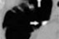
A patient with central HCC tumors showed the most striking tumor-to-parenchyma contrast at 5 to 10 seconds, Kanematsu said, displaying the corresponding image. "Note that the proximal hepatic artery is intensely enhanced, and the trunk enhancement weaker at (5 to 10 seconds)," he said. "At 15 seconds, the HCC is mildly enhanced, but liver parenchyma has also enhanced, so that contrast between tumor and liver is reduced, which indicates that this timing is great for the hepatic arterial phase. At 35 seconds the liver parenchyma is intensely enhanced, and the HCC is clearly washed out, indicating that this timing is optimal for the portal venous phase."
For detecting hypervascular HCC, the optimal scan delay for the hepatic arterial phase after a 30-second contrast injection was between 5 and 10 seconds, he concluded. Optimal timing in the portal venous phase was 35 seconds.
The study provides important baseline information on the chosen protocol, though Kanematsu acknowledged in response to a question after his talk that not all patients would be well served by a fixed scan delay; those with severely reduced cardiac output could require substantially increased scan delays.
Higher iodine concentrations improve lesion conspicuity
Another study presented during the RSNA session, by Dr. Renate Hammerstingl and colleagues from Frankfurt University Hospital in Germany, concluded that high concentrations of iodinated contrast (e.g., 90 mL of iomeprol 400) improved the demarcation and delineation of hepatic tumors compared with the same overall iodine dose at lower concentrations (74 HU versus 64 and 66 for iomeprol 300 and 350, respectively, p = 0.01). Similar findings regarding high iodine concentrations at a constant dose have previously been demonstrated for other anatomic regions.
Saline injection prolongs useful scan window
In a third RSNA study, Dr. Takamichi Murakami from the Osaka University Graduate School of Medicine in Osaka, Japan, presented his group's research aimed at optimizing scan timing in double arterial-phase imaging after the arrival of contrast material to the abdominal aorta. In the study, 60 patients were randomized to one of four groups that received different doses of contrast with and without a saline chaser.
Early arterial-phase images showed good arterial enhancement beginning at about 10 seconds after the arrival of contrast medium, the group concluded, with late arterial-phase images optimized at about 17 seconds after contrast arrival, using a fixed injection duration of 25 to 30 seconds and a fixed amount of contrast material at 1.7-2.0 mL/kg. The use of a saline chaser both increased maximum arterial enhancement and prolonged the useful scan period, according to Murakami.
"Hepatocellular carcinoma showed peak enhancement about 7 seconds after maximum aortic peak enhancement," the group wrote in an accompanying abstract.
By Eric Barnes
AuntMinnie.com staff writer
December 28 2004
Related Reading
CT tracks liver involvement in HTT, February 20, 2004
Korean investigators share angiographic pitfalls of HCC, March 29, 2003
For hypovascular liver tumors, late-phase CT is the way to go, August 18, 2003
All-in-one CT protocol improves evaluation of potential living liver donors, August 12, 2002
Optimizing CT protocols for liver lesions, July 23, 2001
Copyright © 2004 AuntMinnie.com