Just as PACS technology has radically changed the expectations of clinicians for access to diagnostic images, and rapid access to patient images has forced radiology departments to improve report turnaround time, the increasing use of multidetector-row CT (MDCT) for cardiac imaging is mandating the adoption of 3D advanced visualization software.
This is requiring both radiologists and cardiologists to learn new workstation software and develop new interpretation skill sets, and has spawned the creation of dedicated 3D laboratories operated by 3D technologist specialists. It has changed workflow interpretation processes in both cardiology and radiology departments. Clinical demands for faster processing, more automation of manual processes, and greater precision is driving 3D software development, especially for cardiac imaging applications.
Advanced visualization software could even change the decades-old tug-of-war between radiologists and cardiologists over control of imaging technology. In the future, those who control the 3D workstation may be the ones who control cardiac CT imaging.
The necessity of 3D
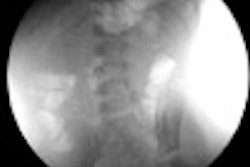
Cardiac imaging using a multidetector-row scanner comes at the price of generating datasets of thousands of images. Multiphase coronary studies can generate 3,000 to 4,000 slices per exam. Facilitating the display and providing a new perspective for expedient review of these datasets require the use of cardiac applications-specific 3D software.
Radiologists and cardiologists alike face the challenge of viewing data acquired in a single plane utilizing multiple other planes. 3D advanced visualization software provides a road map to efficiently manage and interpret the data using volumetric analysis without becoming overwhelmed by it.
Examples of 3D utilization
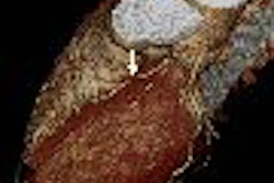
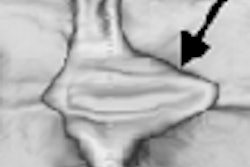
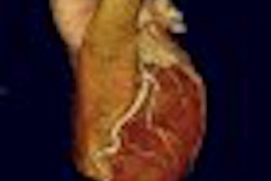
Volume renderings are useful for evaluating complex anatomy, such as coronary artery anomalies and fistulas, and for some cardiologists and radiologists, they help define the axial images for immediate review. They also contribute to the diagnostic evaluation, but are of greatest value in displaying clinical anatomy for cardiologists, cardiovascular surgeons, referring physicians, and patients.
"Radiologists are accustomed to creating 3D anatomic images in their minds from viewing a 2D dataset," said Dr. Robert Falk, a radiologist at Jewish Hospital of Louisville, KY. "A 3D image created through volume rendering provides that image in a glance, and facilitates concentration on the axial images in a large dataset that are the most important."
"3D is not yet a standalone diagnostic function. 3D volume rendering (3DVR) is a time-saving device," he added. "If I am looking at a coronary CTA (CT angiography) on a patient who has had a bypass, the first thing that I am going to do is 3DVR because it gives me the global picture of where the bypasses are going. 3DVR alleviates the need for me to go back and forth through hundreds of images to create this same global picture in my mind."
Dr. John Rumberger, Ph.D., medical director of the Healthwise Wellness Diagnostic Center in Dublin, OH, almost exclusively uses maximum intensity projection (MIP) and volume rendering to interpret coronary CTA procedures. He alternates between both presentation types in a rapid and convenient manner when viewing a coronary segment or a larger portion of the cardiac anatomy.
"MIP can provide valuable information about the condition of a vessel by enabling the interpreting physician to present the (coronary CTA) data in an appropriate context," Rumberger said. "But because it does not encode depth relationships in a single projection image, I also use volume rendering."
Dr. Jeffrey Hellinger, director of cardiovascular imaging of Children's Hospital of Philadelphia in Pennsylvania, also stresses that using the 3D structural overview techniques of MIP and VR, combined with 2D MIPs and curved planar reformations (CPRs), provides the most efficient interrogation methods to study cardiac structural morphology, coronary arteries and veins, thoracic and abdominal aorta, and peripheral medium and small arteries.
Formal clinical studies have not yet been published to prove that volumetric imaging improves cardiac surgeons' performance and clinical patient outcomes, but numerous cardiac specialists anecdotally state that such images are of great assistance. The ability of 4D imaging to graphically show how valves open and close is one such example often referenced in conferences.
Plaque measurement
3D is starting to be utilized to measure plaque. Plaque tends to accumulate within the wall of a vessel, causing the lumen to shrink. In some cases, the vessel wall with plaque may be pushed out, and not necessarily narrow the lumen diameter.
Volumetric rendering dramatically shows areas of concern. A future generation of 3D software is expected to provide comparative analysis, becoming an important clinical tool to identify asymptomatic patients who require aggressive drug therapy.
Assessing bypass graft and stent patency
Cardiac CT is useful for assessing bypass graft patency, and can detect occlusions and stenoses of bypass grafts with very high accuracy. A similar utilization is assessing stent patency. However, the high-density metallic nature of stents can create artifacts that prevent the evaluation of in-stent restenosis.
3D, particularly volume rendering, is also useful for diagnosing coronary artery anomalies, because it images the arteries so well. 3D volumetric rendering can show the problem clearly and enables a radiologist to select the portion of axial images that are the most relevant.
Current limitations
Spatial resolution limits the ability of CTA to provide exact quantitative measures of stenosis. Advanced visualization software provides a stenosis percentage, but its accuracy is limited by the image itself.
The effect of calcium blooming can result in overestimation of the degree of stenosis, creating false-positive results.
The unpredictable heart rhythm of a patient with atrial fibrillation, ectopic beats, or erratic heart rates makes imaging and 3D analysis very difficult, even with the use of beta-blockers. Motion artifacts diminish the quality of 2D- and 3D-rendered images. Adaptive segmented reconstruction algorithms, however, can help minimize motion artifacts and reduce banding.
Accurate quantification of the severity of coronary stenosis is limited or inaccurate for vessels with a diameter of less than 4 mm because most software was initially designed for use with larger values, such as the aorta.
Tasks that require manual activities now must be automated to keep the interpretation of a cardiac CT study from being too time-consuming. Even with current automation, performing measurements is regarded as too time-consuming and cumbersome.
Dr. Elliot Fishman, director of diagnostic radiology and body CT at Johns Hopkins Hospital in Baltimore, recommends that "artificial intelligence be perfected for the calculation of ventricular volumes and ejection fractions by enabling a user to simply touch the appropriate chambers on a workstation screen, rather than requiring the drawing of lines to define endocardial borders."
Workflow
Models of workflow to preprocess, reconstruct, and utilize advanced visualization software for 3D rendering are highly individualistic within radiology and cardiology departments. Despite significant improvements by 3D software and CT modality vendors, processing data is still considered time-consuming.
The quality of volumetric images depends on the reconstruction of high-quality image datasets. Reducing dataset overlapping can substantially reduce the amount of data to process. This is now feasible with 64-slice MDCT-generated images.
Selection of reconstruction filters will affect both image quality and data volume being processed and moved through a network. Ideally, consensus should be reached on the algorithms used. The importance of establishing a comprehensive protocol that defines the parameters to acquire, reconstruct, postprocess, and archive data is essential to achieve a smooth workflow.
3D cardiac imaging workflow is highly individualized. Some radiologists and cardiologists prefer to generate advanced visualization imaging themselves without assistance by a 3D specialist. Others exclusively depend upon dedicated 3D labs for this work. In many hospitals, technologists process a significant portion of the data, configuring it to facilitate additional software utilization by interpreting clinicians.
At least one independent outsourcing service for 3D processing is in operation. 3DR of Louisville, KY, offers comprehensive dedicated 3D lab services targeted to community hospitals. The company offers 3D processing for routine and stat cases on a 24/7 basis, according to Michael Lillig, 3DR CEO.
The need for multidepartment and hospital-wide access to image processing is making the use of dedicated 3D processing workstations less popular. Not only are such dedicated workstations very expensive, but access is limited by their availability and even location in a hospital.
Thin client-server models, providing image access and manipulation capability using a back-end high-performance computer and 3D-rendering server, have proliferated. These "enterprise" models allow users at PACS workstations with 3D software installed, for example, to deliver commands to server hardware, which then reconstruct 3D volume-rendered images in real-time and send them for viewing to the requesting workstations. Most central servers have storage ability to retain three to 12 months of thin-slice data.
Retention of 3D images and axial images within a 4,000-slice dataset is also an individual determination by a hospital. The network infrastructure of a PACS can be severely compromised by the routine transfer of more than 1,000 image datasets, and permanent archiving of raw data may be an unnecessary use of archival and backup storage. Protocols developed by a radiology or cardiology department need to define what reconstructed images should be retained, along with the created volumetric images.
Many existing PACS networks are being replaced with gigabyte switches to prevent any delays in transferring MDCT images from the scanner to designated enterprise servers or dedicated workstations.
Turf battles and training
Could 3D software give radiologists a chance to retain some control of cardiac CT before it slips away to cardiologists? Some industry observers have posited this scenario.
Dr. Tony DeFrance, an interventional cardiologist and director of the Cardiovascular CT Angiography Education Center in San Francisco, runs a "boot camp" for level 1 and 2 training in the use of a 3D workstation. Cardiologists and radiologists attend the hands-on course together.
"Cardiologists have a strong understanding of coronary and cardiac anatomy and the best way to integrate diagnostic procedures with patient management," he said. "Radiologists know more about CT operation, protocols, image acquisition, reconstruction, and contrast administration."
"The key to proficiency is to be the person actually operating the 3D workstation, reviewing cases and interactively manipulating the data," he continued. "It is my experience that 80% of CCTA is expertise with the 3D workstation. The other 20% is image acquisition expertise and didactic knowledge."
At DeFrance's course, cardiologists and radiologists are forced to work for 40 to 50 hours together in a five-day time span. They end up better recognizing the strengths and contributions of the other's discipline. "By the end of the course, radiologists and cardiologists are helping each other and collaborating," he said. "My hope is that this experience will provide a model for professional collaboration in cardiac treatment centers."
According to DeFrance, once advanced visualization skills are mastered, review of a CTA study averages 15 minutes. "Becoming competent is highly individualized. The requirements of the ACR (American College of Radiology) and the ACC (American College of Cardiology) are guidelines that do not guarantee proficiency."
Future trends
Dedicated workstations accessible only within 3D labs and CT imaging suites are heading for obsolescence. Thin client, Web access by PCs to high-performance enterprise servers, which will provide all the processing capability of today's 3D dedicated workstation simultaneously for a much larger number of concurrent users, is presumed by modality vendors and independent software providers alike.
Mark Traffas, cardiovascular business manager of Vital Images of Minnetonka, MN, anticipates the seamless integration of advanced visualization data with PACS, RIS, and HIS to users, allowing reports and images to be transferred wherever needed. An industry objective is to develop easier real-time report creation tools than those that currently exist, he noted.
A major R&D challenge to software developers is the intelligent integration of cardiac imaging from multiple modalities and other diagnostic sources to present to individual specialists exactly what they need in the format of preference, according to Colin Roberts, a member of the cardiac imaging product analysis team at Barco of Kortrijk, Belgium. He noted that software developers face the challenge of needing to become clinically astute to better understand customized workflow requirements.
Another anticipated near-future trend is that individual logins may dictate the images to be initially transmitted to a physician, minimizing and optimizing network traffic. A specific referring physician may only need volumetric images and a handful of axial ones.
Similarly, different levels and functions of automated image processing, based on physician preference and department protocols, will be conducted in the background by powerful systems utilizing artificial intelligence.
Steve Sandy, vice president of marketing for TeraRecon of San Mateo, CA, said that TeraRecon's new iNtuition platform architecture, introduced at RSNA 2006, is designed to blur the difference between 3D dedicated workstations and 3D enterprise imaging solutions by completing much of the editing work in advance based on individual customer preferences using preset filters. He believes that this will provide an immediate enhancement of efficiency to users.
The collective goal of developers of advanced visualization software is to develop programs that analyze, understand, and anticipate individual physicians' work patterns and automatically generate exactly what is needed, where and when it is needed. Ideally, the ultimate automation of 3D labs would make the required mastery of 3D processing commands by radiologists and cardiologists obsolete skills for all concerned.
By Cynthia Keen
AuntMinnie.com contributing writer
March 28, 2007
Disclosure notice: Ms. Keen occasionally performs consulting work with several medical imaging vendors.
Related Reading
Survey finds routine use of 3D visualization, December 18, 2006
3D reveals what axial images can't in small-bowel CT, November 15, 2006
Requirements for continued advancement of 3D applications, November 8, 2006
Part II: Medical image processing has room to grow, September 4, 2006
3D penetrates trauma imaging niche, August 24, 2006
Copyright © 2007 AuntMinnie.com