Heart failure due to diastolic dysfunction is the most common cause of heart failure in patients with normal left ventricular ejection fraction (LVEF), researchers agree. But there is debate when it comes to the best methods of diagnosis.
A sample of this divergence can be found in a recent editorial in the Journal of American College of Cardiology, whose authors refute a previously stated view that diagnostic heart failure (DHF) cannot readily be diagnosed by echocardiography.
On the contrary, Dr. Jae K. Oh and colleagues assert in their "counterviewpoint" article, DHF is reliably characterized on 2D and Doppler echo by the presence of abnormal myocardial relaxation, decreased compliance, and increased filling pressure in the setting of normal LV dimensions when LVEF is normal on 2D echo (JACC, February 7, 2006, Vol. 47:3, pp. 500-506).
According to Oh et al, the viewpoint paper by Mauer's group that sparked the rebuttal advanced the position that "1) Doppler-derived diastolic parameters do not provide information on intrinsic passive diastolic properties, thus, diastolic function cannot be diagnosed by Doppler echocardiography; and 2) because delayed relaxation and/or stiffened passive properties may not be the unifying pathophysiologic mechanisms in all patients who represent with heart failure and normal ejection fraction, the term 'heart failure with normal ejection fraction (HFNEF)' is preferred over the use of the term 'diastolic heart failures (DHF)' to describe these patients" (JACC, 2004, Vol. 44, pp. 1543-1549).
Taking on the first point first, Oh et al wrote that the Mauer team's cogently written contention was that Doppler echocardiography mainly reflects diastolic filling pressure rather than intrinsic diastolic properties of the heart. "They further suggested that DHF should be reserved for heart failure caused by an abnormality of intrinsic diastolic function of the heart defined by slowed relaxation or an upward shift of the LV end-diastolic pressure-volume relation (EDVPR)," Oh et al wrote of the paper.
Determining diastolic property and filling pressure with echo
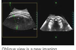
Comprehensive echocardiography uses several techniques including M-mode, 2D, Doppler, color-flow imaging, and myocardial (tissue Doppler) imaging in the context of clinical findings, Oh et al explained.
Thus, "left ventricular relaxation is assessed by isovolumetric relaxation time (IVRT), mitral inflow Doppler velocities, tissue Doppler measurement of mitral annular early diastolic velocity (Ea), and color M-mode measurement of mitral inflow propagation velocity (Vp)," Oh et al wrote. "As myocardial relaxation becomes normal, the rate of decrease in the LV pressure is slower, and IVRT becomes longer, but LV filling pressure is not necessarily elevated. If LV filling pressure is not elevated, the E velocity of mitral inflow decreases and deceleration time (DT) is prolonged."
Patients with normal myocardial relaxation at rest do not develop abnormally increased filling pressure as a result of stress or exercise, Oh et al wrote. Reduction and delay in Ea velocity of the mitral annulus, reduced early diastolic thinning rate on the LV posterior wall, or early diastolic mitral annular motion on M-mode echo can all be used to identify slowed myocardial relaxation.
"In healthy individuals, longitudinal motion of the mitral annulus (Ea) increases with exercise or increased early diastolic filling," they wrote. "However, in individuals with abnormal relaxation and reduced Ea, Ea is less influenced by loading conditions. When myocardial relaxation is markedly abnormal, an early mitral diastolic flow velocity with rapid deceleration is followed by an increased mid-diastolic flow velocity as LV mid-diastolic pressure falls with continuing relaxation."
LV pressure increases abnormally with a moderate decrease in compliance, and transmural filling during atrial contraction is "detected as a shortening of the mitral A-wave duration, whereas the duration of the flow reversal in the pulmonary vein during atrial contraction becomes longer than the mitral A duration," they wrote.
The authors also took issue with Mauer et al's statement that these measurements are elaborate, difficult to obtain, and not routinely performed. Oh and colleagues asserted the following:
- Mitral inflow velocity is and should be recorded routinely.
- Pulmonary venous recording can also be obtained in most patients, though it is technically challenging.
- These measurements represent not a "surrogate index of diastolic function," but rather an abnormal increase in LV pressure with atrial contraction, thus decreased compliance.
- With decreased compliance, LV pressure occurs earlier in diastole, shortening DT and IVRT and increasing mitral E velocity.
- Filling pressure can be estimated by echocardiography in most patients.
- In the setting of reduced ejection fraction, mitral inflow DT correlates well with capillary Wedge pressure.
Mauer et al also argued against use of the term DHF to describe patients with a normal LVEF who have symptoms and signs of heart failure, in favor of calling it heart failure with normal ejection fraction, according to Oh et al.
Heart failure with normal ejection fraction includes many nonmyocardial and myocardial abnormalities that can cause heart failure with normal ejection fraction, Oh et al noted. However, DHF accurately denotes those patients who present with symptoms of heart failure, normal ejection fraction, increased filling pressure, and evidence of diastolic dysfunction, but no other nonmyocardial abnormalities that can cause heart failure.
"In patients with clinical evidence of heart failure, normal (ejection fraction) on 2D echocardiography immediately suggests the potential diagnosis of DHF," they wrote. "Doppler, color-flow imaging, and myocardial tissue imaging can confirm or exclude the diagnosis of DHF by assessing intrinsic diastolic function and estimating diastolic filling pressure."
Detecting diastolic dysfunction in asymptomatic patients may offer an opportunity to manage the underlying etiology and prevent progression to DHF, they concluded.
By Eric Barnes
AuntMinnie.com staff writer
February 28, 2006
Related Reading
US in the ICU: An essential tool in critical care treatment, February 3, 2006
Heart disease in women often eludes angiography, February 2, 2006
Heart disease spotted, treated too little in women, September 2, 2005
Hypertrophic cardiomyopathy seems underrecognized in women, August 29, 2005
Echocardiography bests treadmill test for diagnosis of CAD in women, June 14, 2000
Copyright © 2006 AuntMinnie.com