Thyroid nodules can be risk stratified based on the number of suspicious features on ultrasound, allowing for an effective reporting and data system for thyroid imaging, according to researchers from Yonsei University in Seoul, South Korea.
In a study of more than 1,600 patients with thyroid nodules, the team found a significant association of malignancy with thyroid features detected by ultrasound, including solid component, hypoechogenicity, marked hypoechogenicity, microlobulated or irregular margins, microcalcifications, and taller-than-wide shape.
The group also found that the probability and risk of malignancy escalated as the number of suspicious features increased, allowing for these markers to serve as the basis for a thyroid imaging reporting and data system (TIRADS).
"Risk stratification of thyroid malignancy according to the number of suspicious US features allows for a practical and convenient TIRADS," the researchers wrote.
While widespread use of ultrasound has led to increased detection of thyroid nodules, it can be difficult to decide which nodules need further evaluation, according to senior author Dr. Eun-Kyung Kim, PhD.
"There have been some papers regarding risk evaluation of thyroid nodules based on ultrasound features, but they are complex [systems]," Kim told AuntMinnie.com. "So we wanted to develop a practical approach to categorize thyroid nodules according to [their] malignant risk."
At their institution, 3,674 ultrasound-guided fine-needle aspiration biopsies were performed on 3,674 focal thyroid nodules in 3,414 consecutive patients between May and December 2008. Patients were included in the study if they had the following:
- Maximal nodule diameter ≥ 1 cm
- Benign or malignant results at cytology
- Thyroid surgery after specimens from cytologic examination classified as suspicious for papillary thyroid carcinoma, indeterminate, or inadequate
In all, 1,658 thyroid nodules in 1,638 patients were included in the study (Radiology, July 19, 2011).
Patients ranged in age from 11 to 81 years, with a mean age of 50.6. Mean nodule size in the study was 19.9 mm, with a range of 10 to 80 mm. Thyroid ultrasound was performed using an iU22 ultrasound scanner (Philips Healthcare), with a 5-12 MHz linear-array transducer; both compound and conventional imaging were performed in all patients.
One of seven radiologists, including three fellows and four radiologists with five to 13 years of experience, performed the exams and described the nodules according to their internal component, echogenicity, margins, evidence of calcifications, and shape at the time of biopsy. Ultrasound-guided biopsy was performed after ultrasound evaluation of the thyroid gland by the same radiologist, according to the authors.
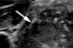
Of the 1,658 nodules, 1,383 were benign and 275 were malignant. From univariate analysis, the researchers found that the ultrasound features of a solid component, hypoechogenicity, marked hypoechogenicity, microlobulated or irregular margins, microcalcifications, and taller-than-wide shape were significantly associated with malignancy.
Multivariate analysis then showed that the risk of malignancy increased with increasing numbers of suspicious ultrasound features.
Thyroid nodule malignancy risk
|
"With these findings, we created TIRADS category 3 (no suspicious US features), 4a (one suspicious US feature), 4b (two suspicious US features), 4c (three or four suspicious US features), and 5 (five suspicious US features) using the risk of malignancy from the BI-RADS categorization," the authors wrote.
The low fitted probability of malignancy for thyroid nodules with no suspicious ultrasound may indicate that it is safe to follow up the mass rather than go to biopsy, according to the authors. However, nodules classified as TIRADS 4 or 5 (at least one suspicious ultrasound feature), are candidates for biopsy.
"This new TIRADS can be easily applied in the clinical field because it is not difficult for those who perform US to count the number of suspicious US features," the authors wrote.
Kim noted that the results were from a single institution, so more studies from other facilities will need to be performed to validate the thyroid imaging reporting and data system.