A significant percentage of thyroid nodule fine-needle aspiration (FNA) biopsies lead to benign findings. But use of thyroid elastography may avoid the need for biopsy in many of these patients, according to research published in the April issue of the Journal of Ultrasound in Medicine.
A research team led by Dr. Manjiri Dighe of the University of Washington Medical Center in Seattle found that quantitative measurements generated from ultrasound elastography could be used to reliably characterize a substantial amount of benign nodules. As a result, only nodules with suspicious findings would go on to receive FNA biopsy.
"With that approach, we may be able to decrease the number of FNA biopsies by 60%," Dighe told AuntMinnie.com. "The ability to confidently diagnose a benign nodule is the most important benefit of elastography."
The researchers sought to determine if a systolic thyroid stiffness index (STSI) -- generated from ultrasound elastography performed by using carotid pulsation as a compression source -- could serve as a pre-FNA biopsy screening tool. They gathered ultrasound data previously acquired from 62 thyroid nodules in 59 patients who had received a thyroid FNA (J Ultrasound Med, April 2010, Vol. 29:4, pp. 565-574).
The ultrasound elastography studies were performed prior to the FNA procedure using a Hi Vision 5500 ultrasound scanner (Hitachi Medical Systems America, Twinsburg, OH) with a 7.5-MHz linear-array transducer. Ten patients were excluded from the study due to inadequate FNA sample results, and one was left out due to inadequate elastography data acquisition.
The sonographers who acquired the studies had more than 10 years of ultrasound scanning experience, as well as more than two years of experience in acquiring elastographic data, according to the researchers. Ultrasound data were processed offline using an angular strain method for elastography; about 200 strain images were generated.
The group then calculated an STSI value by dividing the strain value near the carotid artery at systole by the corresponding strain value in the nodule. A higher STSI value suggests a stiffer thyroid and an increased likelihood of malignancy.
The nodules were classified into two FNA screening categories based on the STSI value. The first category (type I) would receive observation only and no FNA biopsy, and the second category (type II) would undergo biopsy.
The researchers determined the cutoff STSI value to differentiate the nodules based on receiver operator characteristics (ROC) analysis.
At 18.43 ± 5.99, the mean STSI for the 12 malignant nodules was significantly higher than the mean index of 6.82 ± 3.54 for the 39 benign nodules (p = 0.00002). The area under the ROC curve for diagnosing papillary carcinoma was 0.942 for distinguishing between malignant and benign nodules.
From the ROC analysis, the researchers found that a cutoff index value of 10 yielded 100% sensitivity, 79.5% specificity, 60% positive predictive value, and 100% negative predictive value.
 |
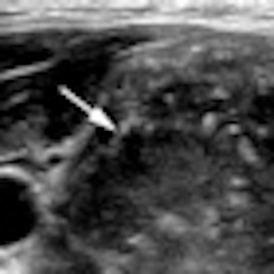
| Left, malignant hypoechoic solid nodule with central areas of calcification and an STSI of 20.1. Right, benign hypoechoic nodule with peripheral calcifications and an index value of 5.6. Images courtesy of the Journal of Ultrasound in Medicine. |
Based on the researcher's guidelines, 60.8% of the biopsies could have been avoided.
"Only 20 (8 benign and 12 malignant) nodules that were classified as type II would have undergone an FNA," the authors wrote. "Thus, by using thyroid ultrasound elastography as a triage tool, it would be possible to limit FNAs to only type II (high probability of malignancy) nodules, thereby decreasing the percentage of benign nodules being referred for an FNA."
The authors noted that future prospective studies will be needed to confirm elastography's efficacy as a triage tool to FNA.
By Erik L. Ridley
AuntMinnie.com staff writer
April 21, 2010
Related Reading
New ultrasound techniques show promise in thyroid nodules, February 5, 2010
Risk of thyroid cancer increased in childhood cancer survivors, October 28, 2009
ARRS study: US can help avoid thyroid biopsy, April 23, 2009
Ultrasound elastography shows potential in thyroid nodules, February 11, 2009
Screening for thyroid cancer worthwhile in some childhood cancer survivors, December 29, 2008
Copyright © 2010 AuntMinnie.com