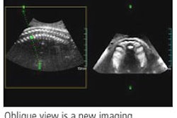
The combination of spatiotemporal image correlation (STIC) and tomographic ultrasound imaging (TUI) may help simplify the fetal echocardiography exam, according to research published in the August issue of the Journal of Ultrasound in Medicine.
"The three-vessel and trachea view, the four-chamber view, and both outflow tracts can be simultaneously visualized using a novel algorithm combining spatiotemporal image correlation and TUI," wrote researchers from the National Institutes of Health in Bethesda, MD; Wayne State University in Detroit; and William Beaumont Hospital in Royal Oak, MI (JUM, August 2006, Vol. 25:8, pp. 947-956).
To evaluate the role of an algorithm combining the two techniques, the researchers examined 237 volume datasets from fetuses with (14) and without (138) congenital heart defects (CHDs). Examinations were performed with STIC using a Voluson 730 Expert system (GE Healthcare, Chalfont St. Giles, U.K.) and hybrid mechanical and curved-array transducers.
Acquisition time ranged from 7.5 to 15 seconds, and the angle of acquisition ranged between 20° and 40°, depending on fetal motion and gestational age. Patients were examined between 14 and 41 weeks of gestation, with a median of 24 3/7 weeks.
After removal of patient identifiers, the exams were retrospectively reviewed offline using version 5.0 of GE's 4DView software, according to the researchers.
A normal four-chamber view was viewed in 99% (193/195) of the volume datasets in patients without heart defects, while a longitudinal view of the ductal arch was visualized in 96% (189/195) of the sets. The three-vessel and trachea view was visualized in 98.5% (192/195) of the sets, while the left outflow tract and short axis were visualized in 93.3% (182/195) and 87.2% (170/195) of the sets, respectively.
The researchers noted that the visualization rates of these planes were higher in fetuses between 20 and 30 weeks of gestational age than in those at less than 20 weeks and those at greater than 30 weeks. These differences, however, were only statistically significant for visualization of the five-chamber view, according to the authors.
For fetuses with heart defects, a four-chamber view was visualized in 85% (17/20) of the volume datasets, while a five-chamber view was seen in 80% (16/20) of the sets. The three-vessel and trachea view was visualized in 65% (13/20) of the sets, while the left outflow tract and short axis were viewed in 55% (11/20) and 70% (14/20) of the sets, respectively.
The researchers noted that simultaneous visualization of the three-vessel and trachea view, the four-chamber view, and both outflow tracts were obtained in 78% (152/195) of the volume datasets from fetuses without heart defects and in 40% (8/20) of those from fetuses with heart defects.
Among fetuses without heart defects, the visualization rates of the three-vessel and trachea view and simultaneous visualization of the three-vessel and trachea view, the four-chamber view, and both outflow tracts were significantly higher when using volume datasets obtained with color Doppler imaging than when using volume datasets obtained with B-mode imaging, according to the study team.
The lower visualization rates of the ultrasonographic planes used in fetal echocardiography among fetuses with heart defects may reflect abnormal spatial relationships, the authors stated.
"The introduction of new display modalities such as the TUI coupled with novel algorithms, such as the one proposed herein, may further simplify the examination of the fetal heart and could reduce the operator dependency that characterizes 2D fetal echocardiography," the authors concluded. "The inability to obtain standard planes with this or previously reported algorithms should raise the possibility of an underlying heart defect. Prospective studies are required to determine the ability of these algorithms as screening methods for the identification of CHDs."
By Erik L. Ridley
AuntMinnie.com staff writer
August 8, 2006
Related Reading
3D brings opportunities to sonographers, August 4, 2006
2D ultrasound offers similar info to 3D/4D obstetric US, June 8, 2006
3D ultrasound brings efficiency gains, May 25, 2006
3D sonography of the endometrium adds value, May 5, 2006
3D ultrasound predicts fetal pulmonary hypoplasia, April 21, 2006
Copyright © 2006 AuntMinnie.com


