A study of nearly 20,000 chest CT scans found that using a commercially available AI tool for pulmonary nodule assessment reduced radiologist reporting time by 14.6 percent, from 21.3 minutes to 18.2 minutes per scan, while also potentially reducing institutional workload by approximately half a full-time radiologist equivalent annually.
- AI chest CT analysis reduced median reporting time by 14.6%, dropping from 21.3 minutes to 18.2 minutes per scan
- Study analyzed 39,323 chest CT examinations across one year before and after AI deployment at Erasmus Medical Center
- Largest efficiency gains occurred for gated thoracic examinations (−41.1% reduction) and thoracic radiologists (−25.0% reduction)
- At typical institutional volumes of 20,000-22,000 annual scans, AI deployment could reduce radiologist workload by approximately 0.5 full-time equivalent
- Emergency department scans showed increased reporting time (7.1%), suggesting AI impact varies by clinical context
Chest CT scans read with a commercially available AI tool for pulmonary nodule assessment reduced reporting times by almost 15%, according to a study published September 1 in Radiology.
A group from Erasmus Medical Center in Rotterdam, the Netherlands, compared radiologist reporting times for one full year before and one full year after deploying a commercially available AI device, with findings suggesting the technology can meaningfully reduce reporting burden in routine clinical practice.
"Chest CT has become a primary tool for identifying pulmonary nodules, yet interpreting these scans remains time-intensive and cognitively demanding, even for experienced radiologists," the researchers wrote. "With increasing imaging volumes and growing pressure on radiology departments, efficiency and accuracy in lung nodule detection and analysis are more important than ever."
The effect of AI on radiologist reporting times has remained an area of ongoing debate, according to the authors. While AI is widely expected to reduce interpretation time, concerns have been raised that additional workflow steps, such as reviewing AI-generated suggestions or verifying flagged findings, could actually slow reporting instead, they explained.
To shed light on the issue in a "real-world" clinical setting, the group conducted a retrospective analysis of chest CT examinations performed at Erasmus Medical Center between September 2021 and May 2024. The final sample comprised 19,433 patients (39,323 examinations: 19,190 pre-AI and 20,133 post-AI). The primary outcome was radiology reporting time, defined as the interval from initial examination opening in the PACS to final report authorization.
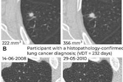
 Example of AI-assisted pulmonary nodule detection and longitudinal volumetric assessment in a 65-year-old woman with peripheral arterial disease, a 24-pack-year smoking history, emphysema, and a family history of lung cancer. An incidental 8.5-mm right lower lobe pulmonary nodule is identified at CT angiography of the abdomen in July 2022 but does not receive follow-up. Subsequent imaging demonstrates interval growth, and CT-guided biopsy confirms stage IA lung adenocarcinoma. Contrast-enhanced axial chest CT images obtained on February 2, 2024, before robotic-assisted thoracoscopic segmentectomy, show a 12.1-mm spiculated right lower lobe pulmonary nodule. (A) Conventional contrast-enhanced axial chest CT image without AI assistance shows a 12.1-mm spiculated right lower lobe pulmonary nodule. (B) Corresponding CT image with AI-assisted pulmonary nodule detection, automated segmentation, and quantitative nodule assessment identifies a solid pulmonary nodule (nodule #1) measuring 13 × 8 (11) mm, with a volume of 739 mm3, a 45% volumetric increase compared with the previous contrast-enhanced chest CT performed on October 9, 2023 (510 mm3), and an estimated volume doubling time (VDT) of 216 days. (C) AI-generated longitudinal nodule analysis displays automated segmentation of the current (February 2, 2024) and previous (October 9, 2023) CT examinations, 3D nodule reconstruction, and quantitative volumetric assessment for longitudinal follow-up. The AI interface is displayed in the original software language (Dutch). RSNA
Further, at institutional scan volumes (approximately 20,000 to 22,000 chest CT exams annually), exploratory modeling suggested an approximate reduction of 0.5 full-time equivalent radiologist workload, the researchers reported.
"Implementation of commercial AI-assisted pulmonary nodule assessment on chest CT scans reduced radiologist reporting time in a real-world clinical setting," the group wrote.
Future studies should assess generalizability across different health care settings and determine whether efficiency gains occur alongside maintained or improved diagnostic performance, the authors concluded.
In an accompanying editorial, Tae Iwasawa, MD, of Yokohama City University in Japan, noted that the study provides compelling real-world evidence that AI can create valuable time within the reporting workflow.
"The next challenge is not simply to read more examinations with fewer radiologists but to use that time to deliver richer, more quantitative, and more clinically meaningful reports," according to Iwasawa.
Ultimately, the success of AI in radiology will be measured not by how many minutes it saves, but by how much it improves patient care, she wrote.
The full study is available here.