A PET/CT-based radiomics approach could help clinicians distinguish between malignant and benign vertebral compression fractures (VCFs), according to a study published October 11 in EJNMMI Research.
Researchers led by Xun Wang, MD, PhD, of Jining Medical University in Shandong, China, developed a radiomics model based on features extracted from F-18 FDG-PET/CT images from a large group of patients. Ultimately, they suggest the model could improve outcomes for patients.
“Early and accurate differential diagnosis of benign and malignant VCFs can allow clinicians to effectively choose appropriate treatment plans and potentially provide improved outcomes,” the group wrote.
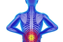
VCFs are small breaks in the vertebrae in the spine and imaging plays a crucial role in their diagnosis, the study authors wrote. Benign VCFs are caused by osteoporosis or trauma, while malignant VCFs may be caused by tumors, such as metastatic solid tumors, myeloma, or lymphoma, they noted.
While CT scans are effective for detecting anatomical signs of both types, when these signs are absent, the diagnosis can be challenging, they added. Conversely, F-18 FDG-PET/CT scans are a common approach for imaging cancer and can provide information about both morphological changes and metabolic status of the diseased vertebra.
In this study, the researchers aimed to combine FDG-PET/CT features and CT features in a radiomics model that could help clinicians make treatment decisions in these cases.
The researchers analyzed imaging from 439 patients diagnosed with vertebral compression fractures after PET/CT scans at their hospital from January 2016 to January 2023. They extracted 26 features from the imaging: nine PET features and 17 CT features, as well as eight clinical variables, such as age, maximum standard uptake value (SUVmax) of FDG radiotracer, osteolytic destruction, and fracture line, for instance.
Using these, the group constructed a combined clinical-radiomics model and a model based on the clinical features alone. They then tested the models on PET/CT images from 44 patients with 20 benign VCFs and 24 malignant VCFs. Performance was based on the area under the curve (AUC) of the models for distinguishing between benign and malignant VCFs.
According to the findings, the AUC of the clinical-radiomics model was 0.948, compared with 0.858 for the clinical model, the authors wrote.
“Combining clinical parameters with radiomics scores based on F-18 FDG-PET/CT can be used to predict the malignancy of vertebral compression fractures with high diagnostic accuracy,” the group wrote.
To date, the diagnosis of the causes of VCFs by CT, as well as other approaches such as MRI, bone SPECT/CT, and PET/CT is largely dependent on the experience of diagnostic physicians, the researchers noted. Adding radiomics to the analysis can provide objective information that is difficult for human eyes to quantify, they suggested.
“We believe that radiomics-based models can improve the diagnostic accuracy and efficiency of diagnostic physicians because radiomics models use mathematical algorithms to describe lesions more objectively and can complement radiologists,” the group concluded.
The full article is available here.