A group in Japan has proposed a method for assessing vertebral compression fractures on x-rays in the absence of MRI – importantly, the method involves elevating patients' heads, but it does not worsen their pain, according to a recent study.
A group led by Keisuke Tsuruta, MD, of Minaminara General Medical Center in Yoshino, found that lateral x-rays taken with patients in supine and 30° head-elevated positions can accurately diagnose acute vertebral compression fractures (AVCFs) and that patients undergoing the imaging reported no pain.
"Our proposed method provides a simple and accurate diagnosis of vertebral compression fractures by using two lateral radiographs and could contribute to alleviating crowded emergency departments," the group wrote, in an article published September 7 in Acute Medicine and Surgery.
Current guidelines recommend an early MRI exam to exclude new spine fractures. However, MRI may be unavailable in certain medical settings or contraindicated because of factors such as claustrophobia or metallic implants, the authors wrote.
An alternative is to compare supine and sitting/standing position x-rays. Yet acquiring these images in patients with acute vertebral compression fractures is challenging, given that patients may require emergency transport and are in severe pain, they added. Moreover, bed rest without elevating the head beyond 30° is recommended in these patients.
"Because MRI is not always immediately available for emergency patients with suspected AVCFs at our facility, the patients first undergo supine and 30° head-elevated lateral radiographs," the group wrote.
In this study, the group aimed for the first time to validate the approach. They retrospectively examined 30 patients with AVCFs who were transported by ambulance to their emergency department between June 2018 and May 2019.
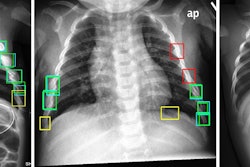
For patients with difficulty in standing or sitting because of severe back pain, x-rays were routinely performed with a 30° head elevation (the patient's head was raised by 30° according to a scale on a stretcher when the radiographs were taken in lateral view). The radiographs were taken centering on the thoracolumbar junction without changing the positions of tube or cassette, the researchers added.
To evaluate the x-rays for fractures, the researchers manually measured vertebral wedging ratios. These measurements indicate how much vertebrae are compressed and are typically expressed as percentages, with higher percentages indicating potentially worse compression fractures. MRIs interpreted by a radiologist served as the final diagnosis.
Out of the 30 patients, a total of 176 vertebrae were included (32 with fractures and 144 nonfractured). The wedging ratios in fractured vertebrae ranged between 5.1% and 24.4%, whereas ratios in nonfractured vertebrae ranged between -6.7% and 4.3%, according to the findings.
Ultimately, the median wedge ratio of fractured vertebrae was significantly higher than nonfractured vertebrae (12.6% vs. -0.5%, p < 0.001) and no patients reported pain during head-elevated positioning, according to the group.
"Comparison of lateral radiographs in supine and 30° head-elevated positions can accurately diagnose AVCF, without aggravating lower back pain," the authors concluded.
The full article is available here.