A new algorithm for automatically segmenting image data delivers fast and reproducible volumes of a wide variety of tumors, satisfying a significant unmet need in neuro-oncology, according to a new study in Academic Radiology. A panel of readers gave the algorithm high marks for how well it approximated manual data segmentation methods.
In the study, researchers developed a semiautomatic segmentation method and used it to segment data from a wide variety of brain tumors, including glioblastomas, meningiomas, and brain metastases. Quantitative validation based on comparing semiautomatic with manual segmentations showed a high level of reproducibility and concordance.
"The ... methodology addresses this unmet clinical need for a segmentation technique that is robust to variation in image quality and tumor type," wrote lead author Bilwaj Gaonkar, from the University of Pennsylvania, and colleagues (Acad Radiol, May 2015, Vol. 22:5 pp. 653-661).
A long quest
The quest for automated brain tumor segmentation has received a lot of attention over the past decade, with dozens of studies proposing different methodologies. However, "despite a substantial body of literature, a method that can segment different types of brain tumors in a clinical setting has remained elusive," the authors wrote.
Moreover, many proposed segmentation methods require specialized preprocessing, and computation times are often so long as to preclude efficient clinical use, they noted. Finally, many algorithms are specific to certain types of tumors and do not generalize well to the variety of tumors found in neuroimaging.
Current methods based on 2D imaging, such as the Macdonald criteria for gliomas or Response Evaluation Criteria in Solid Tumors (RECIST) for general oncology, offer a rough estimate of tumor volume, but accurate volumetry requires complete segmentation, leading to the search for a fast and accurate automated technique.
Early methods based on a technique called "fuzzy clustering" led to large numbers of false-positive voxels being labeled as tumors. Later techniques based on level sets and active contours often failed when tumors were aggressive and structurally complex. More recently, machine learning techniques have done better, but they are often quite tumor-specific and are very sensitive to changes in noise and imaging protocol.
The current study proposed a semiautomated method for complete lesion segmentation for applications in neuro-oncology. The tumor segmentation technique is "semiautomatic, fast, and based on a relatively simple learning-free algorithm," Gaonkar and colleagues wrote. It remains accurate in the face of noise, scanner variation, processing variation, and tumor heterogeneity.
The technique relies on the adaptive geodesic algorithm described by Gaonkar and Shu to segment brain tumors, a method originally devised to segment the vertebral column on CT images using adaptive geodesic distance, the authors wrote. Geodesic refers to the generalization of straight lines to curved spaces.
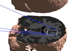
In the algorithm, a seed region is placed inside the tumor and the segmentation algorithm is initiated. The adaptive geodesic distance, a mathematical measure that can be computed at any image voxel, provides joint quantification of the spatial distance of the voxel from the seed region and the variation of the image intensity profile between the voxel and the seed -- "both of which are important clues for tumor segmentation," the authors explained.
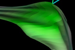
The algorithm computes adaptive geodesic distance at every image voxel to create an adaptive geodesic distance transform image, which appears as the geodesic distance-weighted inverse of the original MR image. Thresholding of this image generates the final segmentation mask.
The retrospective study used a random sample of cases available on the university's PACS network. Gaonkar and colleagues applied their algorithm to the segmentation of 54 brain tumors, including 24 glioblastomas, 15 meningiomas, and 15 brain metastases. The MRI data varied widely in terms of acquisition protocol, resolution, and pixel spacing. Some patients were scanned on 3-tesla MRI systems from GE Healthcare and Siemens Healthcare, while others were imaged on 1.5-tesla scanners, the study team wrote.
Qualitative validation of the images was based on physician ratings provided by three clinical experts, while quantitative validation was based on comparing semiautomatic and manual segmentations. On a scale of 1 to 5, a score of 5 meant that the algorithm performed perfectly in data segmentation.
| Average ratings of algorithm performance | |||
| Tumor type | Rater 1 | Rater 2 | Rater 3 |
| Glioblastoma | 4.3 | 4.2 | 3.7 |
| Meningioma | 4.6 | 4.5 | 4.7 |
| Metastasis | 4.4 | 4.0 | 4.2 |
"The slightly [lower] values for glioblastomas are consistent with the fact that these tumors are heterogeneous and extremely challenging to segment, manually or semiautomatically," Gaonkar and colleagues noted. "Meningiomas, on the other hand, are relatively homogeneous and therefore comparatively easier to segment."
The main drawback of the method is the dependence of segmentation on manual initialization, they wrote. An experienced clinician must provide a small set of initial voxels that drive subsequent computation. Multifocal tumors require multiple initializations, and the technique does not work when there are motion artifacts.
As for technique, volumetric analysis has repeatedly been shown to outperform 1D or 2D methods, and semiautomated techniques have been shown to be more reproducible, according to the authors. The new method represents a "significant advancement over currently available segmentation tools available to clinicians," they wrote. Furthermore, it is computationally efficient, does not require any special computer hardware or preprocessing, and is insensitive to scanner parameters.
"Our analysis demonstrates that this methodology produced segmentation results that are on par with a manual segmentation performed by a radiologist," Gaonkar and colleagues concluded.
"Being able to accurately assess tumor volume has important implications for both the diagnosis and management of patients with brain malignancies," they wrote. "The outlined methodology addresses this unmet clinical need for a segmentation technique that is robust to variation in image quality and tumor type."


