Hands-on intensive echocardiogram training with lectures and one-on-one discussion to build the complete cardiac ultrasound protocol.
Echocardiography | The Hands-On Cardiac Ultrasound Imaging and Doppler Course
Aug 13th, 2017Aug 17th, 2017
Dallas, TX
US
Phone:972-353-3200, 800-845-3484
Latest in Home

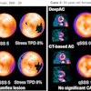
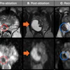


![(A-C) Representative whole-body maximum-intensity projection images and regional fused PET/CT images from three histologically confirmed osteosarcoma patients who underwent paired [68Ga]Ga-B7-H3-BCH PET/CT and 18F-FDGE PET/CT within seven days. (D) Multimodal imaging evaluation of Patient Three, including x-ray, MRI (T2-weighted imaging, T2WI), CT, and B7-H3 PET/CT.](https://img.auntminnie.com/mindful/smg/workspaces/default/uploads/2026/05/mei.XUQJWkpAJI.jpg?auto=format%2Ccompress&dpr=2&fit=crop&h=167&q=70&w=250)


