This course will combine instructor presentations and interactive sessions with the opportunity to examine your communication skills, medicolegal issues, and real case scenarios in which you will be the judge.
Mammography: Take This Job and Love It
Jul 25th, 2014
Latest in Home


Sponsored
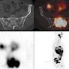
Webinar: AI for CT & PET/CT Imaging
June 4, 2026


