The most practical and effective protocol to image and assess the vessels in the upper abdomen. Learn how to document every pertinent feature of each organ. Detailed assessment of AAA and renal vascular disease. 24-hour scan lab.
Hands-On Abdominal Ultrasound Imaging
Dec 16th, 2014Dec 19th, 2014
Latest in Home


Sponsored
Webinar: AI for CT & PET/CT Imaging
June 4, 2026
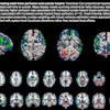
Brain SPECT offers insight into suicide risk
June 5, 2026


