This course has been designed specifically for those involved in gathering clinical data to support regulatory approval and marketing of medical devices. With the recent amendments to the Medical Device Directives there are more stringent requirements to ensure the quality of the data submitted for CE marking of devices, and the data that needs to be generated once the device is on the market.
Medical Device Clinical Investigations and Evaluations
Mar 14th, 2010Mar 15th, 2010
Latest in Home


Sponsored
Webinar: AI for CT & PET/CT Imaging
June 4, 2026
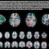
Brain SPECT offers insight into suicide risk
June 5, 2026


