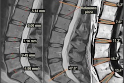
A standardized MRI classification system developed by researchers significantly improves the accuracy and consistency of lumbar spinal stenosis grading among radiologists and physicians, with overall correctness increasing and interobserver reliability improving after implementing the structured educational intervention.
- Overall correctness improved from 54.5% to 61.2% after implementing the standardized classification system among 37 physicians across six specialties.
- Interobserver reliability increased from 0.71 to 0.75, meaning radiologists and physicians showed better agreement in their assessments.
- Cases discrepant by more than one grade decreased from 3.7% to 2.0%, reducing significant diagnostic disagreements.
Classification systems could help improve correct MRI analysis on lumbar spinal stenosis, according to research published July 4 in Academic Radiology.
Researchers led by Pamela Walsh, MD, from Hofstra University in Hempstead, NY highlighted success from its standardized MRI lumbar spinal stenosis classification system, which they reported significantly improved grading concordance and interobserver reliability among radiologists and other physicians.
“We recommend implementing a consistent reporting language… to improve multidisciplinary communication, research, quality improvement, and optimal patient management,” Walsh and colleagues wrote.
MRI is the preferred method for evaluating lumbar spinal stenosis. However, grading the severity of stenosis has been subjective and inconsistent among radiologists and non-radiology physicians who treat spine pathologies. The researchers pointed out the lack of a standardized reporting system for lumbar spinal stenosis.
The Walsh team developed its own structured educational intervention using a classification system. They studied this intervention’s impact on correctness and interobserver reliability of MRI grading among radiology and non-radiology spine physicians.
The researchers based their classification system used for the educational intervention on the Lee classification system, though with modifications. These included assessing compression of the thecal sac in any direction, the addition of criteria that the thecal sac was compressed up to the level of the cauda equina nerve roots, and providing more detail in assessing the cauda equina nerve roots, using a half/50% cutoff for mild and moderate categories.
The study analyzed 114 lumbar disc levels. And 37 physicians across six specialties graded stenosis in pre-intervention (routine practice) and post-intervention phases following training with the classification system. The specialties included the following: musculoskeletal (MSK) radiology, neuroradiology, radiology residents, orthopedic surgery, neurosurgery, and physiatry.
The classification system led to overall correctness and interobserver reliability improving, as well as cases discrepant by more than one grade decreasing.
Impact of educational intervention on MRI lumbar spinal stenosis grading | |||
Measure | Pre-intervention | Post-intervention | P value |
Overall correctness | 54.5% | 61.2% | 0.006 |
Interobserver reliability | 0.71 | 0.75 | 0.03 |
Cases discrepant more than one grade | 3.7% | 2.0% | 0.002 |
During the pre-intervention phase, MSK radiologists had the highest percentage of cases correct (67.7%) and lowest discrepant cases (1.6%). Post-intervention, neuroradiologists had the highest correct (64.9%) and lowest discrepant (0.7%).
Finally, MSK radiologists had the highest intraclass correlation coefficient values in both pre-intervention (0.774) and post-intervention (0.781) phases.
The study authors suggested their intervention could provide a framework for standardizing lumbar spinal stenosis assessment at other institutions.
“Future research can explore AI algorithms trained with a standardized classification system agreed upon by a large multidisciplinary group, such as in this study, to improve diagnostic accuracy and efficiency as well as integrate clinical data to establish clinical utility of these models,” they added.
Read the full study here.