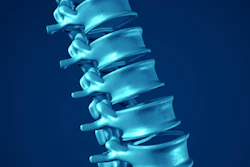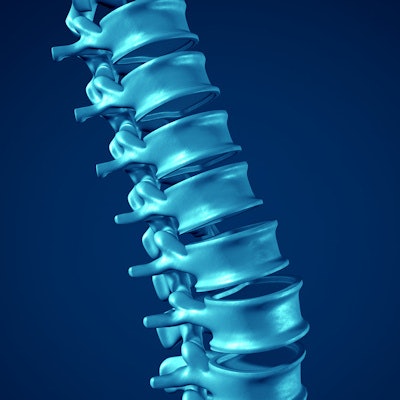
Fast low-angle shot (FLASH) MRI can significantly improve the detection of degenerative lumbar spine disease, according to research presented at the annual meeting of the American Roentgen Ray Society in Honolulu, HI.
In a session on musculoskeletal imaging, Dr. Karthik Shyam of Ganga Medical Center and Hospital in Coimbatore, India, told attendees that adding a FLASH sequence to standard MRI protocols can improve the detection rates in some cases by more than 85%.
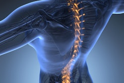
The incidence of lumbar degenerative spine disease is increasing worldwide, and in progressively younger populations, Shyam noted. While T1- and T2-weighted MRI imaging is used routinely in these patients, it is limited in its ability to detect soft tissue pathology (disk herniations) and small osseous bone changes involved in the disease, he said.
Importantly, the inability to visualize small osseous changes in patients can complicate surgery and result in inadequate clearance of calcification, Shyam added.
FLASH sequences (3D T1-weighted gradient-recalled echo sequences) are optimized parameters for MRI scanners that provide improved bone/soft tissue signal differences. Applying a grayscale inversion to these sequences results in images that appear similar to contrast-enhanced CT -- a gold standard approach, yet one not indicated for all patients due to the risk of radiation exposure, Shyam explained.
Shyam and colleagues conducted a study that added FLASH sequences to routine T1- and T2-weighted MRI scans in 148 patients with lower back pain. The aim was to see if the proposed protocol could detect osseous changes not seen on routine MRI. They used conventional CT imaging at the site of interest as a reference, with two experienced radiologists analyzing the images.
"The results are quite promising," Shyam said.
Specifically, improvements were seen in detecting foraminal osteophytes, corner bony endplate defects, and vacuum phenomena. He noted the addition of FLASH sequences to routine MRI improved the detection of annular calcification by 86%.

Ultimately, the protocol could help screen patients who likely may need an additional CT scan to evaluate the extent of the disease, as well as aid novice readers in identifying osseous changes, Shyam suggested.
"Addition of FLASH enables detection of clinically significant osseous changes in degenerative lumbar disease," he concluded.