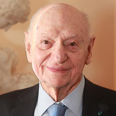
Dr. Alexander Margulis, who chaired the department of radiology at the University of California, San Francisco (UCSF) from the 1960s to the 1980s, has died, according to a post on the university's website. He was 97.
Margulis was one of the giants of radiology, taking the reins at UCSF in 1963, at a time when plain-film radiography was essentially the only medical imaging modality. He led the department until 1989, along the way helping to foster an explosion of imaging technologies such as ultrasound, nuclear medicine, interventional radiology, CT, MRI, PET, and more. Under his leadership, UCSF became one of the premier radiology residency programs in the world.
Dr. Alexander Margulis.Born in 1921 in Belgrade, Serbia, Margulis was a medical student when Germany invaded the former Yugoslavia in 1941. He and his family escaped the country in 1944 on a refugee ship from Naples, Italy. After their arrival in the U.S., he and his brother were admitted to Harvard Medical School, according to the university's post.
He completed his residency in diagnostic radiology at the University of Michigan and in 1954 joined the faculty of the University of Minnesota. He then became a U.S. citizen and served in the U.S. Army Medical Corps at Fort Bragg, NC. Margulis later joined the faculty at Washington University in St. Louis.
Margulis was recruited to be the chair of UCSF's department of radiology in 1963, a position he held for the next 26 years, seeing both the rapid growth of the department and of radiology. He trained and mentored many radiologists who went on to leadership positions at other academic institutions, and he played key roles in the development of medical imaging modalities such as MRI and molecular imaging.
For example, under Margulis' initiative, UCSF in 1971 bought the second head CT scanner acquired by an academic institution in the U.S., an EMI system that had a price tag in today's dollars of $3.5 million. UCSF also played a major role in the development of superconducting MRI scanners; in fact, if it hadn't been for Margulis, MRI might still be called "nuclear magnetic resonance," a term that was discarded as the modality neared clinical use. Other important contributions under his watch were made in obstetric ultrasound.
Margulis stepped down as UCSF's department head in 1989, but he continued to be affiliated with the university in various roles. UCSF named a professorship after him, and it created the Margulis Society in 1991 as a fundraising organization for UCSF alumni and faculty. The society provides financial support to UCSF residency teaching programs, and it also pays for university residents to attend the annual RSNA meeting.
In 2000, Margulis became a clinical professor of radiology at Weill Cornell Medical College of Cornell University, after his wife, radiologist Dr. Hedvig Hricak, PhD, became chair of radiology at Memorial Sloan Kettering Cancer Center in New York City.
One of Margulis' most long-lasting achievements for UCSF was his role in selecting a second campus for the university in San Francisco. Working with city officials, he steered the development away from San Francisco's suburbs to an area on the southern edge of the city's downtown, in a neighborhood now known as Mission Bay. The area continues to grow and develop and is where many of UCSF's radiology activities are located.
"Alex touched the lives and careers of innumerable radiologists and scientists and he enriched us all with his humor, leadership, and sagacious support of imaging science. ... His impact on the health of individuals everywhere and the careers of myriad radiologists in the United States and abroad cannot be overstated," UCSF wrote in the post.


![Examples of ultrasound findings and techniques. (A) Images in a 39-year-old male patient with a mass in the left thigh. The mass is heterogeneous on the B-mode US image (compared with the patient in D) and showed increased microvascularity (superb microvascular imaging [SMI]) and shear-wave elastography (SWE) values. Undifferentiated pleomorphic sarcoma was diagnosed at biopsy (with pleomorphic rhabdomyosarcoma in surgical specimen). (B) Images in an 18-year-old male patient with a mass in the left leg. The mass is hypoechoic on the B-mode image, with no other findings suggestive of malignancy. The lesion is in contact with the cortex of the tibia, which is slightly irregular. CT revealed a doubtful anteromedial tibial erosion. The microvascular study demonstrated high vascularization, suggestive of malignancy. Periosteal Ewing sarcoma was diagnosed with both histologic and immunohistochemical confirmation. (C) Images in a 69-year-old female patient with a lump growing on the outside of the left leg. Multiple SWE examinations were performed (please note the high values obtained in the measurements, whereas the color map highlights the stiffness relative to adjacent tissues). SMI showed areas of increased vascularization to target for sampling. Undifferentiated spindle cell sarcoma was diagnosed at biopsy, with residual leiomyosarcoma in the surgical specimen after neoadjuvant therapy. (D) Images in a 56-year-old female patient with a mass in the right thigh. The mass is heterogeneous at both B-mode ultrasound (similar to patient A) and MRI (coronal T2-weighted spectral attenuated inversion recovery [SPAIR]; T1-weighted pre-contrast and postcontrast imaging), which even shows uptake after the administration of paramagnetic contrast material, which is traditionally suggestive of malignancy. Low values at SMI and elastography are suggestive of benignity. Spindle cell lipoma was diagnosed at biopsy, with atypical spindle cell lipomatous tumor in the surgical specimen.](https://img.auntminnie.com/mindful/smg/workspaces/default/uploads/2026/08/images-radiol250278fig2.APCFLSvX6p.jpg?auto=format%2Ccompress&fit=crop&h=100&q=70&w=100)


![Examples of ultrasound findings and techniques. (A) Images in a 39-year-old male patient with a mass in the left thigh. The mass is heterogeneous on the B-mode US image (compared with the patient in D) and showed increased microvascularity (superb microvascular imaging [SMI]) and shear-wave elastography (SWE) values. Undifferentiated pleomorphic sarcoma was diagnosed at biopsy (with pleomorphic rhabdomyosarcoma in surgical specimen). (B) Images in an 18-year-old male patient with a mass in the left leg. The mass is hypoechoic on the B-mode image, with no other findings suggestive of malignancy. The lesion is in contact with the cortex of the tibia, which is slightly irregular. CT revealed a doubtful anteromedial tibial erosion. The microvascular study demonstrated high vascularization, suggestive of malignancy. Periosteal Ewing sarcoma was diagnosed with both histologic and immunohistochemical confirmation. (C) Images in a 69-year-old female patient with a lump growing on the outside of the left leg. Multiple SWE examinations were performed (please note the high values obtained in the measurements, whereas the color map highlights the stiffness relative to adjacent tissues). SMI showed areas of increased vascularization to target for sampling. Undifferentiated spindle cell sarcoma was diagnosed at biopsy, with residual leiomyosarcoma in the surgical specimen after neoadjuvant therapy. (D) Images in a 56-year-old female patient with a mass in the right thigh. The mass is heterogeneous at both B-mode ultrasound (similar to patient A) and MRI (coronal T2-weighted spectral attenuated inversion recovery [SPAIR]; T1-weighted pre-contrast and postcontrast imaging), which even shows uptake after the administration of paramagnetic contrast material, which is traditionally suggestive of malignancy. Low values at SMI and elastography are suggestive of benignity. Spindle cell lipoma was diagnosed at biopsy, with atypical spindle cell lipomatous tumor in the surgical specimen.](https://img.auntminnie.com/mindful/smg/workspaces/default/uploads/2026/08/images-radiol250278fig2.APCFLSvX6p.jpg?auto=format%2Ccompress&fit=crop&h=112&q=70&w=112)