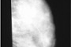
SAN DIEGO - Accurate breast biopsies can prevent future work-up of benign lesions, and play a crucial role in presurgical localization and neoadjuvant chemotherapy planning. At the American Roentgen Ray Society meeting on Monday, a multi-institutional group discussed a new breast biopsy marking device that is visible on sonography and mammography.
"The indications for using this biopsy device are to demonstrate using the mammographic and sonographic correlation in a lesion that may not be well visualized, or may not be visualized, on the mammogram (only)," said Dr. Terese Kaske from the Sally Jobe Breast Center in Englewood, CO. Kaske’s co-authors are based at Sally Jobe, as well as Park Nicollet in Minneapolis; the Women’s Imaging Center in Denver; Sacred Heart Hospital in Allentown, PA; and Cedar Valley Medical Center in Waterloo, IA.
For this study, 11 physicians at the five centers used the UltraCor device (SenoRx, Aliso Viejo, CA) to place biopsy markers in 43 sites in 39 patients. The device consists of a 10-cm needle with depth markers placed every 1 cm, and a beveled applicator containing the biopsy site marker. The marker is made up of five resorbable pellets, which are delivered to the site by depressing a syringe-type plunger.
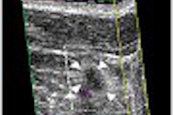
The pellets are made of polylactic and polyglycolic acids. Carbon dioxide within each pellet allows for visibility on ultrasound; a stainless steel wire embedded in the first pellet is visible only on a mammogram.
The study sought to assess functional characteristics and marker visibility. Ultrasound images were obtained at deployment and at a follow-up exam up to five weeks after biopsy. The overall visibility on ultrasound was scored on a 3-point scale, with zero for not visible and 2 for highly visible. The participants used a standardized form to record the data.
In the majority of cases (41), the marker was placed after 14-gauge biopsy; after fine-needle aspiration in one case; and after one directional vacuum-assisted biopsy procedure. An average of five samples were collected per case.
According to the results, the UltraCor device applicator was placed independently in 88% of the cases and with coaxial needle guidance in 12%. The operators rated the beveled needle easy or satisfactory to direct to the biopsy site in 98% of the cases. Pellet deployment force was rated easy in all cases, with no difficult or failed deployments reported.
Finally, the investigators found that the marker was visible on ultrasound at deployment in 95% of the cases, and visible in 90% of the cases with completed follow-up (to date, 30 patients), five weeks after biopsy. Cases in which the marker placement was not visible on mammographic follow-up occurred when the pellets were deployed far medially or far laterally to the lesion, Kaske said.
"The device is easy to use although it requires a little bit of finesse, which is quickly learned," she concluded. For example, in hard lesions, rather than deploying all of the pellets simultaneously, which could send the markers beyond the lesion, Kaske’s group determined that it was more efficient to place one pellet in the lesion and the rest adjacent to it.
In answer to audience questions, Kaske said the group had no reported foreign body reactions to the pellets, and that marker migration was not an issue either. She added that the studies have shown the UltraCor device to be MRI-compatible.
By Shalmali PalAuntMinnie.com staff writer
May 5, 2003
Related Reading
Preop needle biopsy a risk factor for infection after breast surgery, April 11, 2003
U.K. researchers explore reasons for biopsy clip movement, March 8, 2003
New adjunctive breast techniques improve positive biopsy rates, February 25, 2003
Post-biopsy gel marker stays put on mammographic follow-up, December 5, 2002
Breast biopsy technology evolves at a rapid pace, May 16, 2002
Copyright © 2003 AuntMinnie.com