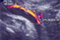
(Radiology Review) A recent retrospective review of sonographically demonstrated muscle atrophy and concomitant rotator cuff pathology was performed to assess their relationship.
The Journal of Ultrasound in Medicine recently published the results. Including an examination of the muscles at ultrasound examination of the rotator cuff adds vital information that may predict the success of rotator cuff surgery, according to Dr. Carolyn M. Sofka and colleagues.
Radiologists at the Hospital for Special Surgery, New York, reviewed 199 ultrasound examinations of the shoulder. Scans were performed using a 7.5 MHz linear array by radiologists trained in musculoskeletal ultrasound. Department protocol included assessment of the rotator cuff tendons and their respective muscles, as well as the long head of the biceps tendon, the deltoid muscle, the subdeltoid bursa, and the acromioclavicular joint. Muscles and tendons were examined in the longitudinal and short-axis planes. Atrophy was diagnosed when the muscle appeared hyperechoic and had decreased bulk.
Muscle atrophy was demonstrated in at least one muscle in 23% of patients. The teres minor muscle was affected in 36% of cases, the infraspinatus in 31%, the supraspinatus in 16%, the biceps brachii in 6%, and the subscapularis in 2%.
"In 34 of the 45 examinations with muscle atrophy, there were concomitant full thickness tendon tears. There were 57 individual tendon tears, with the following distribution: 64% supraspinatus, 38% infraspinatus, 16% long head of biceps, 7% subscapularis and 2% deltoid," they said.
Seventy-seven percent of the patients with supraspinatus muscle atrophy had full-thickness tendon tears, and 60% of patients with infraspinatus muscle atrophy had associated infraspinatus tendon tears. Fatty atrophy was shown in at least one muscle in almost one fourth of shoulders studied.
"Although focus has been maintained primarily on integrity of the rotator cuff tendons, our study suggests that evaluation of the muscles should become a standard component of the comprehensive shoulder sonographic examination because a finding of muscle atrophy may indicate to the examiner the presence of a concomitant pathologic condition such as a rotator cuff tear," they concluded.
Detection of Muscle Atrophy on Routine Sonography of the Shoulder
Carolyn M. Sofka, et al
Department of Radiology and Imaging
Hospital for Special Surgery
535 E 70th St., New York, NY 10021 USA
J Ultrasound Med 2004 (August); 23:1031-1034
By Radiology Review
August 16, 2004
Copyright © 2004 AuntMinnie.com