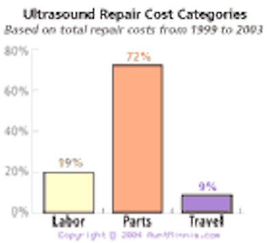
Many healthcare equipment managers have heard that alternative sources for replacing ultrasound transducers cost less than what manufacturers charge, but they don't know how much less. A five-year comparison study shows that costs to replace all types of ultrasound transducers were lower with parts from independent service organizations (ISOs) compared with those from original equipment manufacturers (OEMs).
![]() |
![]() |
According to the study, replacing invasive and noninvasive transducers with ISO parts cost 35% less than using OEM parts, while replacing transesophageal echocardiography (TEE) transducers with ISO parts cost 23% less than using OEM parts.
Additionally, the study concluded that parts replacement represents more than 70% of the total repair budget (See Ultrasound Transducer Replacement Cost Averages chart, above).
- Incorporating certain precautionary measures can reduce overall transducer costs.
- Use a bite guard during an entire procedure when using TEE transducers;
- Properly follow the manufacturer's recommendations on cleaning solutions and length of immersion time;
- Properly package a transducer when shipping for repair to prevent further damage;
- When moving a cart, remove the transducers and move by hand, being careful not to get cables caught in front of wheels, doorknobs, equipment, etc.
Information provided to AuntMinnie.com comes from the GENISYS databank of USCS Equipment Technology Solutions, Brookfield, WI. For more information on asset management visit the USCS Web site at www.us-cs.com, or call 800-558-6377. USCS Equipment Technology Solutions and GENISYS are registered trademarks.







![Examples of ultrasound findings and techniques. (A) Images in a 39-year-old male patient with a mass in the left thigh. The mass is heterogeneous on the B-mode US image (compared with the patient in D) and showed increased microvascularity (superb microvascular imaging [SMI]) and shear-wave elastography (SWE) values. Undifferentiated pleomorphic sarcoma was diagnosed at biopsy (with pleomorphic rhabdomyosarcoma in surgical specimen). (B) Images in an 18-year-old male patient with a mass in the left leg. The mass is hypoechoic on the B-mode image, with no other findings suggestive of malignancy. The lesion is in contact with the cortex of the tibia, which is slightly irregular. CT revealed a doubtful anteromedial tibial erosion. The microvascular study demonstrated high vascularization, suggestive of malignancy. Periosteal Ewing sarcoma was diagnosed with both histologic and immunohistochemical confirmation. (C) Images in a 69-year-old female patient with a lump growing on the outside of the left leg. Multiple SWE examinations were performed (please note the high values obtained in the measurements, whereas the color map highlights the stiffness relative to adjacent tissues). SMI showed areas of increased vascularization to target for sampling. Undifferentiated spindle cell sarcoma was diagnosed at biopsy, with residual leiomyosarcoma in the surgical specimen after neoadjuvant therapy. (D) Images in a 56-year-old female patient with a mass in the right thigh. The mass is heterogeneous at both B-mode ultrasound (similar to patient A) and MRI (coronal T2-weighted spectral attenuated inversion recovery [SPAIR]; T1-weighted pre-contrast and postcontrast imaging), which even shows uptake after the administration of paramagnetic contrast material, which is traditionally suggestive of malignancy. Low values at SMI and elastography are suggestive of benignity. Spindle cell lipoma was diagnosed at biopsy, with atypical spindle cell lipomatous tumor in the surgical specimen.](https://img.auntminnie.com/mindful/smg/workspaces/default/uploads/2026/08/images-radiol250278fig2.APCFLSvX6p.jpg?auto=format%2Ccompress&fit=crop&h=112&q=70&w=112)
