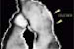
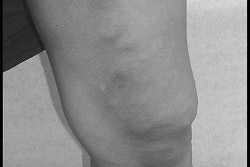
(Ultrasound Review) Deep venous thrombosis (DVT) of the leg is a major cause of pulmonary embolus, which leads to a significant number of deaths each year. Color Doppler ultrasound is the imaging modality of choice for patients suspected of having DVT of the leg.
The American Journal of Roentgenology recently published work by researchers at University of Rochester Medical Center in New York whose objective was to determine whether it was necessary to include the more difficult calf veins in the ultrasound examination when the patient has no calf symptoms.
In a randomized prospective study, performed ultrasound scans were performed using two different protocols. "We compared patient outcomes using two protocols: one routinely and the other selectively evaluating the calves completely during sonographic assessment of the lower extremities in patients with suspected deep venous thrombosis," they reported.
The first group of patients had their thigh, knee and calf veins scanned, while in the second group, only the deep calf veins were evaluated when there were physical signs evident, and only the symptomatic area was evaluated. Patients with a negative result were followed for a 3-month period and a consequent knee or thigh DVT or a pulmonary embolus was considered an adverse outcome. Using a 5-7 MHz linear array, color and spectral Doppler and grayscale were used to assess the deep veins. DVT was considered present when the veins were incompressible on grayscale transverse images, and color and spectral Doppler was used for confirmation.
Results demonstrated no adverse outcome in the first group of 235 patients that included calf vein evaluation. There were two adverse outcomes in the second group of 261 patients at follow-up. "Seven isolated deep venous thrombi of the calf were detected. Four of these occurred in the incomplete calf protocol versus three in the complete calf protocol group," they reported.
They concluded "no significant risk exists in performing a calf examination only if there are symptoms or physical signs in the calf and in not evaluating the calf otherwise. The calf should not be part of the routine leg sonography protocol for suspected deep venous thrombosis unless signs or symptoms are present in the calf."
Randomized prospective study comparing routine versus selective use of sonography of the complete calf in patients with suspected deep venous thrombosisGottlieb, R. H. et al
Department of radiology, University of Rochester Medical Center, Rochester, NY
AJR 2003 January; 180:241-245
By Ultrasound Review
May 5, 2003
Copyright © 2003 AuntMinnie.com