Ultrasound can be an effective screening approach for blunt abdominal trauma (BAT) cases, but more imaging studies may be needed for some high-risk patients, according to a study from the University of California, San Diego.
"In patients with negative ultrasound studies, those with hematuria or fractures of the lower ribs, lumbar spine, or pelvis have a 24 times higher risk of abdominal injury than patients with no risk factors," said Dr. Claude Sirlin, assistant professor of body imaging at the university. "Ultrasound is a screening test, but must be interpreted in the context of clinical and other imaging findings."
The UCSD research team reviewed a database of 4,000 patients screened for BAT at UCSD, a level 1 trauma center, between 1994 and 2001. The group’s goal was to test the hypothesis that patients with hematuria or various fractures have a higher risk of missed abdominal injury after negative ultrasound than patients without such factors.
The group also sought to assess individual risk factors and to propose a new triage algorithm. Sirlin presented the research team's findings at the 2002 RSNA meeting in Chicago.
The study team retrospectively identified all patients who had a negative screening ultrasound, and reviewed the trauma registry to identify those with rib, lumbar spine, or pelvic fractures or with hematuria (more than 50 rbc on urinalysis obtained during resuscitation). The ultrasound was considered positive if free fluid, hematomas, or parenchymal abnormalities were found, and was considered negative otherwise, Sirlin said.
The ultrasound results were then compared with the best available gold standard (CT, repeat ultrasound, peritoneal lavage, surgery, autopsy, or clinical outcome). Of the 3,679 patients with negative ultrasound findings, 494 met the study criteria as patients at high risk of missed injuries.
Of the remaining 3,185 patients considered to be at low risk for missed injury, eight had injuries, four of which required surgery. In the 494 high-risk patients, 30 had injuries, 10 of which required surgery.
Of the 216 rib fractures, 13 had abdominal injuries (three of which needed surgery). Eight of the 105 lumbar spine fracture cases had injuries, of which three needed surgery. Pelvis fracture cases (174) included nine abdominal injuries (two of which needed surgery). The 96 hematuria patients had 15 abdominal injuries, five that needed surgery, Sirlin said.
"Hematuria carried the greatest risk (of the individual risk factors), with one out of six having abdominal injuries and one out of 19 having surgical injuries," he said.
 |
Only 16% (606 of 3,680) patients with negative ultrasound studies would have had unnecessary CT exams, Sirlin said. As a corollary, the UCSD team found that patients with negative ultrasound and no risk factors rarely had missed injuries (one in 398 had abdominal injuries and one in 796 had surgical injuries).
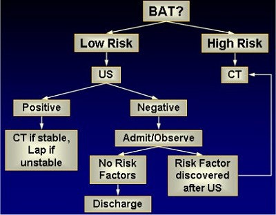
Based on the research, the UCSD researchers created a new triage algorithm. As a result of the triage algorithm, more than 99.8% of patients with an injury would be triaged to CT or laparotomy, with less than 0.2% being discharged from the hospital with a missed injury, Sirlin said.
 |
| Charts courtesy of Dr. Claude Sirlin. |
Based on the group's research, 16 CT scans would be required to find an injury, and 49 for a surgical injury in the high-risk group, compared with 398 and 796, respectively, for the low-risk patient population, he said.
A useful screening study
In a subsequent UCSD presentation, Dr. Michele Brown reported on the institution's experience in ultrasound screening of 4,451 BAT patients between 1994 and 2002. Ultrasound results were compared to findings at diagnostic peritoneal lavage (DPL), CT, repeat ultrasound, cystography, laparotomy, autopsy, and clinical observation.
Screening produced a sensitivity for detection of abdominal injury of 84%, specificity of 97%, positive predictive value of 64%, and negative predictive value of 99%, Brown said, concluding that abdominal ultrasound is a useful screening exam in trauma patients.
"The greatest limitation is the detection of injuries without hemoperitoneum, particularly retroperitoneal and enteric injuries," she said.
By Erik L. RidleyAuntMinnie.com staff writer
May 1, 2003
Related Reading
Imaging technologies reduce unnecessary appendectomies in children, December 13, 2002
Ultrasound differentiates causes of abdominal pain in kids, October 4, 2002
Emergency Imaging of the Acutely Ill or Injured Child, September 10, 2002
SPECT coregistration improves treatment at pediatric care center, June 18, 2002
Ultrasound disappoints in abdominal trauma, December 14, 2001
Copyright © 2003 AuntMinnie.com