Ultrasound-guided endovenous laser treatment of varicose veins is a patient-friendly option for treating a condition that affects about one in five American adults, according to Dr. Robert Min, director of Cornell Vascular and vice chairman of radiology at Weill Medical College of Cornell University in New York City.
Min said that two-year follow-up of 499 limbs treated with diode laser energy for saphenous vein reflux "confirmed a closure rate of 93%. So with this procedure we have a recurrence rate of 7% compared to a 10% to 25% historical recurrence rate with surgical stripping, which was the prior gold standard." Min presented his group's results at the 2003 meeting of the Society of Interventional Radiology in Salt Lake City.
Min, who developed the technique at Cornell Vascular, said the "key to success is careful duplex ultrasound assessment of the vein to confirm diagnosis and to assess the veins. A vein that is extremely tortuous is not a good candidate for this procedure, because catheter access is too difficult. However, that is only a small minority of veins, and those patients will require surgery."
Compared to surgery, which requires general anesthesia, the laser procedure is done under local anesthesia so it can be done on an outpatient basis, he explained. "And cosmetically, this procedure is far superior to surgery because the only scar is the small puncture wound at the venous port. Surgery, on the other hand, leaves significant scars," Min said.
He said that many clinicians consider varicose veins a simple cosmetic problem. But for many patients varicose veins are highly symptomatic, typically causing pain at rest, which can disturb sleep. Generally, the condition is most common in multiparous women, but Min said that it can also affect men, usually in their late 50s.
![]() |
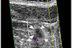
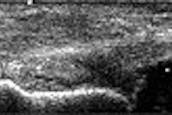
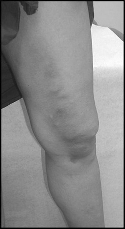
Before (above) and after (below) pictures of a patient who underwent ultrasound-guided endovenous laser treatment. Images courtesy of Dr. Robert Min and SIR.
![]() |
The treatment is performed with an 810-mm diode laser (Diomed, Andover, MA), which is threaded into the greater saphenous vein via a 600-micron laser fiber. Veins are assessed percutaneously under ultrasound guidance. Patients are evaluated clinically with duplex ultrasound at a week, one month, six months, 12 months, and 24 months.
Min said he has had no instances of perforation or laser-associated damage with the procedure, attributing his success to careful ultrasound guidance. "We don’t fire the laser until we are sure that we have reached the target area and we fire as we are backing out of the vein," Min said.
Patients are able to walk immediately after the procedure and most patients return to work within days, he said. Post-procedure patients have minor bruising, and pain that can be treated with over-the-counter analgesics.
Min said about 50 U.S. centers are currently offering the procedure, and he expects that number to double by year’s end.
By Peggy PeckAuntMinnie.com contributing writer
April 25, 2003
Related Reading
Pseudoaneurysm size affects success of thrombin injection with US, April 1, 2003
US-guided needle therapy provides quick relief for joint injuries, December 5, 2002
Coding changes on tap for October 1, 2002, July 16, 2002
Ultrasound guidance keeps steroid injections safe, April 22, 2002
Intervention can help or harm in athletic injuries, February 17, 2002
Copyright © 2003 AuntMinnie.com



![Examples of ultrasound findings and techniques. (A) Images in a 39-year-old male patient with a mass in the left thigh. The mass is heterogeneous on the B-mode US image (compared with the patient in D) and showed increased microvascularity (superb microvascular imaging [SMI]) and shear-wave elastography (SWE) values. Undifferentiated pleomorphic sarcoma was diagnosed at biopsy (with pleomorphic rhabdomyosarcoma in surgical specimen). (B) Images in an 18-year-old male patient with a mass in the left leg. The mass is hypoechoic on the B-mode image, with no other findings suggestive of malignancy. The lesion is in contact with the cortex of the tibia, which is slightly irregular. CT revealed a doubtful anteromedial tibial erosion. The microvascular study demonstrated high vascularization, suggestive of malignancy. Periosteal Ewing sarcoma was diagnosed with both histologic and immunohistochemical confirmation. (C) Images in a 69-year-old female patient with a lump growing on the outside of the left leg. Multiple SWE examinations were performed (please note the high values obtained in the measurements, whereas the color map highlights the stiffness relative to adjacent tissues). SMI showed areas of increased vascularization to target for sampling. Undifferentiated spindle cell sarcoma was diagnosed at biopsy, with residual leiomyosarcoma in the surgical specimen after neoadjuvant therapy. (D) Images in a 56-year-old female patient with a mass in the right thigh. The mass is heterogeneous at both B-mode ultrasound (similar to patient A) and MRI (coronal T2-weighted spectral attenuated inversion recovery [SPAIR]; T1-weighted pre-contrast and postcontrast imaging), which even shows uptake after the administration of paramagnetic contrast material, which is traditionally suggestive of malignancy. Low values at SMI and elastography are suggestive of benignity. Spindle cell lipoma was diagnosed at biopsy, with atypical spindle cell lipomatous tumor in the surgical specimen.](https://img.auntminnie.com/mindful/smg/workspaces/default/uploads/2026/08/images-radiol250278fig2.APCFLSvX6p.jpg?auto=format%2Ccompress&fit=crop&h=100&q=70&w=100)


![Examples of ultrasound findings and techniques. (A) Images in a 39-year-old male patient with a mass in the left thigh. The mass is heterogeneous on the B-mode US image (compared with the patient in D) and showed increased microvascularity (superb microvascular imaging [SMI]) and shear-wave elastography (SWE) values. Undifferentiated pleomorphic sarcoma was diagnosed at biopsy (with pleomorphic rhabdomyosarcoma in surgical specimen). (B) Images in an 18-year-old male patient with a mass in the left leg. The mass is hypoechoic on the B-mode image, with no other findings suggestive of malignancy. The lesion is in contact with the cortex of the tibia, which is slightly irregular. CT revealed a doubtful anteromedial tibial erosion. The microvascular study demonstrated high vascularization, suggestive of malignancy. Periosteal Ewing sarcoma was diagnosed with both histologic and immunohistochemical confirmation. (C) Images in a 69-year-old female patient with a lump growing on the outside of the left leg. Multiple SWE examinations were performed (please note the high values obtained in the measurements, whereas the color map highlights the stiffness relative to adjacent tissues). SMI showed areas of increased vascularization to target for sampling. Undifferentiated spindle cell sarcoma was diagnosed at biopsy, with residual leiomyosarcoma in the surgical specimen after neoadjuvant therapy. (D) Images in a 56-year-old female patient with a mass in the right thigh. The mass is heterogeneous at both B-mode ultrasound (similar to patient A) and MRI (coronal T2-weighted spectral attenuated inversion recovery [SPAIR]; T1-weighted pre-contrast and postcontrast imaging), which even shows uptake after the administration of paramagnetic contrast material, which is traditionally suggestive of malignancy. Low values at SMI and elastography are suggestive of benignity. Spindle cell lipoma was diagnosed at biopsy, with atypical spindle cell lipomatous tumor in the surgical specimen.](https://img.auntminnie.com/mindful/smg/workspaces/default/uploads/2026/08/images-radiol250278fig2.APCFLSvX6p.jpg?auto=format%2Ccompress&fit=crop&h=112&q=70&w=112)