VIENNA - Ultrasound is considered to be a rather challenging modality for detecting ureteral stones, but it worked like a charm for Drs. Jongmin Kim and S. H. Kim from the Jinju Gospel Hospital in Jinju, South Korea. Ultrasound left no stone unfound in the team’s eight-month study of emergency-room patients, as confirmed in follow-up imaging studies and clinical interventions.
"We prospectively evaluated ultrasound, IVP, and sometimes CT in 153 consecutive patients presenting with renal colic for the emergency evaluation of ureteral stones," Dr. Jongmin Kim said yesterday in a presentation at the European Congress of Radiology. "US was performed in a state of moderate bladder distension after ingestion of (500 ml) fluid prior to the study."
The diagnostic criterion for ureteral stones was an echogenic focus having posterior acoustic shadowing within the ureteral lumen, and the final diagnosis was confirmed by additional imaging, clinical follow-up, or urological intervention in all cases, Kim said. For the purposes of the study, ureteral stone locations were divided into upper, middle, and lower thirds of the ureter.
According to the results, ureteral stones (mean size 8.0 mm, range 3.5 to 23.8 mm) were detected in 140/153 cases. The remaining 13 patients were diagnosed with appendicitis (n=3), choleodocholithiasis (n=1), choleocystitis (n=1), acute pyelonephritis (n=1), hemorrhagic ovarian cyst (n=1), diverticulitis (n=1), and five unknown cases.
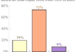
"Ninety-five cases or 68% (of the ureteral stones) were located at the lower third of the ureter, including 69 cases at the ureteropelvic junction," Kim said. "And 29% and 3% were in the upper and middle thirds, respectively."
Ninety-nine cases or 71% revealed calcified stones on follow-up radiography, but the remaining 41 cases did not. In one case, CT failed to visualize a stone seen with ultrasound. Hydronephrosis was noted in 108/140 (77%) of the patients.
"It was difficult for ultrasound to trace throughout the ureter, in many cases due to bowel gas," he said. "Nonetheless, there were some cases where moderate bladder distension could help visualize (stones). Posture change could also help to avoid bowel gas (artifacts) posterior to ureteral stones in many cases."
Anatomic landmarks such as the psoas muscle and ureteropelvic junction, as well as the use of color Doppler ultrasound, were also helpful in differentiating the ureter from vessels, and ureteral stones from bowel gas, he said. All diagnoses were confirmed with IVP, CT, or clinical intervention.
"Ultrasound was very useful for the diagnosis of ureteral stones, with a sensitivity of 100%," Kim concluded. An audience member said the patients seemed quite small and slender on follow-up CT, and asked if their diminutive size might have aided the performance of ultrasound.
"I am Korean," Kim said. "The average weight is not so heavy. But we didn’t exclude anyone from the prospective study."
By Eric BarnesAuntMinnie.com staff writer
March 7, 2004
Related Reading
US calculates splenic volume within ten minutes, January 19, 2004
US combo boosts detection, characterization of perianal inflammation, January 15, 2004
3-D ultrasound shows promise in neonatal, pediatric neurosonography, November 4, 2003
3-D ultrasound offers reliable assessment of gallbladder, bile ducts, May 5, 2003
Power Doppler US spots tumor vascularity for prostate cancer staging, April 30, 2003
Copyright © 2004 AuntMinnie.com