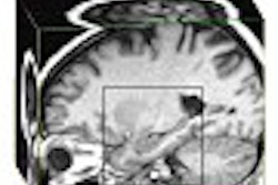
(Radiology Review) MR imaging proved helpful in demonstrating the site and cause of arterial, venous, and nervous compressions in patients with thoracic outlet syndrome (TOS), according to French radiologists at Centre Hospitailer Regional Universitaire de Lille. They compared the MRI findings for symptomatic patients and normal volunteers in a study published in Radiology.
Fifty-four patients with clinically proven TOS and 54 controls underwent MRI, initially with the arm in a neutral position beside the body, and then with the arm hyperabducted 1300 and externally rotated.
"Patients were included in the study when at least two provocative clinical tests reproduced the symptoms," they reported. Control subjects had both sides scanned while patients had the symptomatic side examined.
Using a 1.5-tesla Magnetom Vision scanner (Siemens Medical Solutions, Erlangen, Germany), coronal scout images of the thoracic outlet were followed by T1-weighted spin-echo sequences comprising 16 sections. The scan then was repeated after postural maneuver and the total scan time taken was 30 minutes.
The following measurements were evaluated:
- Interscalene triangle angle
- Maximum thickness of the anterior scalene muscle
- Minimum costoclaviclular distance
- Distance separating the inferior subclavius muscle and the chest wall
- Maximum subclavius muscle thickness
- First rib angle
Retropectoralis minor space measurements included distance separating the posterior pectoralis minor and the axilla at the level of the vessels, as well as the maximum thickness of the pectoralis minor muscle.
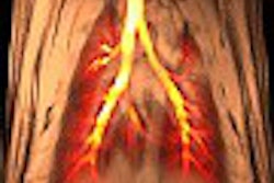
Any compression of the vessels and brachial plexus was noted, as was any compression of the subclavian vein in the prescalene space. Vessels were considered compressed when there was a 30% reduction in arterial diameter and a 50% reduction in venous diameter. They diagnosed nerve compression when the fat surrounding the brachial plexus was absent, or when the brachial plexus was closely adjacent to bone.
Results demonstrated subclavian vein compression in the interscalene triangle in 47% of volunteers, and the authors suggested that venous compression be carefully interpreted given this finding.
"Patients with TOS had a smaller costoclavicular distance after the postural maneuver, a thicker subclavius muscle in both arm positions and a wider retropectoralis minor space after the postural maneuver than did volunteers," they discovered.
Thoracic outlet: assessment with MR imaging in asymptomatic and asymptomatic populationsDemondion, X., et. al.
Department of osteoarticular radiology, Hôpital Roger Salengro, Centre Hospitailer Regional Universitaire de Lille, Lille, France
Radiology 2003 May; 227:461-468
By Radiology Review
August 19, 2003
Copyright © 2003 AuntMinnie.com