MRI can help differentiate between gestational trophoblastic disease and other postpartum or postabortion symptoms, according to a paper in the American Journal of Roentgenology.
"Retained products of conception (occur) with greater frequency after termination of pregnancy...common symptoms include vaginal bleeding and abdominal pelvic pain," wrote lead author Dr. Jaime Noonan from Emory University School of Medicine in Atlanta. "Retained products of conception may be treated conservatively...whereas gestational trophoblastic disease may require chemotherapy" (AJR, August 2003, Vol. 181:2, pp. 435-439).
For this retrospective study, pelvic MRI was performed in four patients who had a histologically confirmed diagnosis of retained products of conception. The exams were performed on a 1.5-tesla whole-body Signa scanner (GE Medical Systems, Waukesha, WI). The MR sequences included axial and sagittal T2-weighted fast spin-echo and contrast-enhanced (0.1 mmol/kg Magnevist, Berlex Imaging, Wayne, NJ), breath-hold T1-weighted in-phase spoiled gradient-echo.
 |
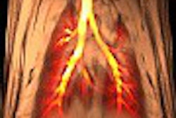
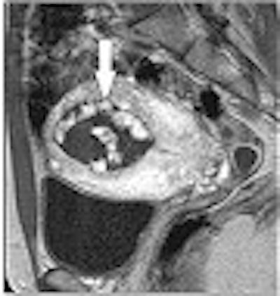
| This 32-year-old woman presented with vaginal bleeding and a serum b -human chorionic gonadotropin level of 45 mlU/mL, two weeks after a spontaneous abortion. An axial T2-weighted image (A) showed an irregular uterine mass (black arrow) of heterogenous intensity. The white arrow points to marked thinning of the myometrium. Image B is from the same patient, a sagittal T1-weighted contrast-enhanced scan that shows areas of brisk enhancement (arrow) in intrauterine mass. Noonan JB, Coakley FV, Qayyum A, Yeh BM, Wu L, Chen L. "MR Imaging of Retained Products of Conception," (AJR, Vol. 181, pp. 435-439). |
"Our results suggest that retained products of conception appear on MR imaging as an intracavitary uterine soft-tissue mass with variable T1 and T2 signal intensities, variable amounts of enhancing tissue, and variable degrees of myometrial thinning and obliteration of the junctional zone," they wrote.
In comparison, previous descriptions of placental-site trophoblastic tumor on MR have suggested that the tumor is intramyometrial in location, rather than intracavitary, they explained.
Of the four women, Patient 3 benefited the most from the MRI, according to co-author Dr. Fergus Coakley, who is chief of abdominal imaging at the University of California, San Francisco.
Patient 3 presented with vaginal bleeding and the passage of tissue two months after treatment of an incomplete spontaneous abortion. Sonography showed an intrauterine mass. Dilatation and curettage had to be terminated prematurely because of persistent bleeding from the cervical os. Because of this, MR was performed and the results showed a 6.1-cm intracavitary mass in the uterus.
"The lesion was isointense to the myometrium on T1-weighted images and heterogeneous on T2-weighted images, and showed focal brisk enhancement after IV administration of gadolinium," the authors reported.
Dilatation and curettage was repeated, with hysteroscopy and laparoscopy showing degenerating chorionic villi without atypical trophoblastic cells. The patient’s symptoms resolved and MRI done two months later showed no uterine mass.
The group acknowledged that limitations of this report included the small patient population and selection bias. In addition, the incidence of retained conception products is quite low (1% of all pregnancies). In Coakley’s department, MR is performed on these patients only when requested, he said.
However, the modality still has value in some patients because of its ability to show anatomic variants in the uterine cavity that may hinder successful treatment.
By Shalmali PalAuntMinnie.com staff writer
August 1, 2003
Related Reading
Cervical ultrasound may predict pre-term birth in women with abortions, July 4, 2003
Uterine fibroid embolization does not impair fertility, April 1, 2003
Copyright © 2003 AuntMinnie.com