A recent neuroimaging study has shown differences in MRI between young people with schizophrenia and those with other mood disorders that have psychotic features. How radiologists can make use of these findings in collaborations with psychiatrists is still unknown, however.
A study presented at the 2002 American Academy of Child and Adolescent Psychiatry (AACAP) in San Francisco found a difference in the gray-matter volume between children and adolescents with schizophrenia and those with mood disorders that had psychotic features.
"These differences suggest that the deficits in gray matter may reflect the underlying neuropathology specific to schizophrenia," said Dr. Mohammed El Sayed, a staff psychiatrist at the University of North Carolina at Chapel Hill. Dr. Linmarie Sikich, an assistant professor of psychiatry at the same institution, collaborated on the study.
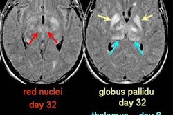
The investigators observed 29 subjects who underwent MRI on a 1.5-tesla Signa scanner (GE Medical Systems, Waukesha, WI). The investigators assessed the patients’ volumes of gray matter, white matter, cerebrospinal fluid, hippocampus, and caudate and lateral ventricles with computer-assisted segmentation. They rescanned 16 of the subjects after they had received 8-23 weeks of antipsychotic medication.
The investigators found that the gray-matter volume was significantly lower in patients with schizophrenia spectrum disorders than in patients with mood disorders, at a significance of less than 0.023. The left hippocampal volume was also significantly smaller in the schizophrenia spectrum group, at a significance of less than 0.025.
"Follow-up scans suggest a slight decrease in the volume of both the left and right hippocampus," Sayed said.
Dr. William Bradley cautioned that the gap between these findings and clinical applicability still exists.
"I am all for a biological basis for psychiatric disease and for favoring a morphologic basis," said Bradley, chair of the American College of Radiology’s commission on neuroradiology and MRI. Bradley is also chair of the radiology department at the University of California, San Diego. "I am not sure that what it means to find less gray matter in schizophrenics." He pointed out that other research has shown that frontal white-matter projections are less myelinated in schizophrenics than in controls.
By Paula MoyerAuntMinnie.com contributing writer
December 16, 2002
Related Reading
MRI findings predict development of symptomatic psychosis, December 11, 2002
In vitro MRI findings support vascular cause of late-life depression, October 18, 2002
Prefrontal cortical activity and dopaminergic function linked in schizophrenia, February 4, 2002
Copyright © 2002 AuntMinnie.com