CHICAGO - Even though there are some cancers that can be seen better with whole-body MRI, researchers at this week’s RSNA meeting said they would prefer to use PET/CT fusion images to stage primary tumors.
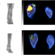
In a study of 50 patients, Dr. Gerald Antoch, a radiologist in the department of diagnostic and interventional radiology at University Hospital Essen in Germany, said that PET/CT found 94 pulmonary lesions in 16 cancer patients compared with MRI, which located 58 lesions in 13 patients.
In eight patients -- 16% of the cohort -- either cervical, thoracic, or abdominal lymph nodes showed pathological FDG uptake as a sign of malignancy, while those tumors were missed on MRI due to their small size of 1 cm or less, Antoch said in his presentation.
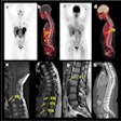
On the other hand, Antoch said that whole-body MRI picked up seven metastases to the bone that were not seen with the PET/CT fusion images. Abdominal parenchymal organs were assessed equally well with both imaging techniques. In the study, Antoch used the Biograph scanner (Siemens Medical Solutions, Erlangen, Germany) for the PET/CT scans; a Siemens 1.5-tesla Sonata MRI system was employed for the comparative scans.
"PET/CT proved superior in detecting lymph node metastases and pulmonary metastases, while MRI was able to more accurately assess metastases to the bone," he said. "Whole-body PET/CT and whole-body MRI examinations, therefore, complement one another in staging of malignant diseases."
But when pressed by session moderator Dr. Peter Conti, professor of radiology at the University of Southern California, Los Angeles, to select one modality to be first used in assessing patients, Antoch said he would go with the PET/CT fusion.
"That’s a good answer," Conti said. "Right now, if I were working up patients for cancer diagnosis, I would use PET/CT to try and answer as many questions as possible. If the patient then complained of back pain and we saw nothing on PET/CT, then I might order MRI or plain-film x-rays."
Conti said using all the tools possible would likely give answers to a lot of questions about the patient, "but the idea is to use one modality to get most of the questions. Right now that looks like using the PET/CT."
In the study, two radiologists read the images produced by the MRI and the PET/CT fusion system; two nuclear medicine physicians also read the PET/CT images. None of the readers were aware of the findings from the other imaging device.
By Edward Susman
AuntMinnie.com contributing writer
December 6, 2002
Copyright © 2002 AuntMinnie.com


