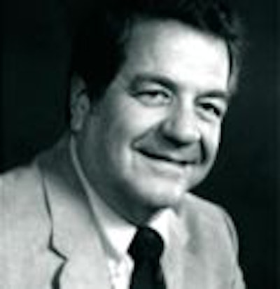
Perhaps no one deserves more credit than Dr. Henry Wagner for the rapid diffusion of the term "molecular imaging" into radiology’s lexicon. Wagner began using the term in the early 1990s, consistently applying it to describe the evolving science of imaging metabolic processes at the molecular level.
In the following question-and-answer session, AuntMinnie.com traces some of Wagner’s thoughts and opinions on molecular imaging.
Wagner is a professor at Johns Hopkins University in the division of radiation health sciences in the department of environmental health sciences at the university’s Bloomberg School of Public Health. He was cofounder in 1959 of the Baltimore institution’s nuclear medicine department with Dr. John McAfee.
Wagner and McAfee developed the first lung scanning procedure for diagnosing pulmonary embolism in the early 1960s. Wagner was a leader in myocardial perfusion research in the 1970s, when his work led to the application of radionuclide angiography to coronary artery disease. In the 1980s, he began studying brain function. Wagner and colleagues at Johns Hopkins produced the first images of dopamine neuroreceptors and opiate receptors in a living person.
Wagner has been widely honored as a writer and educator, and has graduated several hundred residents from his nuclear medicine program. The Society of Nuclear Medicine in Reston, VA, established the Henry Wagner lecture series at its 1996 annual meeting. Wagner's "Highlights" lecture has concluded every SNM annual meeting since 1976.
Where did the term "molecular imaging" come from?
The term arose when it became clear that nuclear medicine could expand greatly the newly developing field of molecular biology. The foundation of medicine extended to a new level of spatial resolution -- from the level of histopathology to the molecular level of DNA and the proteins that RNA produced. Receptors became a focus of medicine and pharmacology.
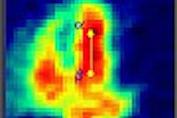
PET and SPECT imaging were beginning to be able to image these receptors using radioligands bound to them. A classic example was the finding by nuclear medicine imaging that the basic "molecular" defect in Parkinson's disease was a great reduction in the dopamine transporter binding of radiotracers in pre-synaptic neurons of basal ganglia.
When was the term "molecular imaging" first used in the imaging community?
I used the term in a Highlights lecture at a Society of Nuclear Medicine meeting. I also wrote an article in the Journal Of Nuclear Medicine, titled "The New Molecular Medicine." In the article, I wrote, "Advances in our understanding of intra- and intercellular communication will revolutionize the practice of medicine" (JNM, January 1993, Vol. 34:1, pp. 165-166).
What exactly does "molecular imaging" refer to? How would you define it?
Again, as I said in the article, "Diagnoses will be molecular, treatment will be molecular, and monitoring the response to treatment will be molecular...Modern scientific medicine is moving away from an organ orientation, and is advancing by leaps and bounds through a holistic approach to disease, exemplified by revolutionary advances in human genetics, immunology, oncology, endocrinology, neuropsychiatry, and molecular biology, all of which are holistic approaches to disease."
Does it incorporate both SPECT and PET?
Yes, both SPECT and PET use molecular "probes" to examine regional biochemistry and function. Each has its own merits. Both will continue to prosper.
Does it include other imaging modalities?
The most immediate extension will be optical imaging, based on the use of fluorescent tracers that will increase spatial resolution by a factor of 1,000. At present this can only be used in small animals and human lesions on the skin or visible by endoscopy. Problems of attenuation and scatter are being solved so that the applications will grow. Ultrasound can measure regional function, but not biochemistry, while the spatial resolution of magnetic resonance spectroscopy is far too low.
Does it include other non-imaging technologies?
Yes, molecular imaging will include studies of blood and other body fluids, in addition to specimens removed at surgery or biopsy.
What are the most exciting areas of molecular imaging research?
The brain is number one, followed by oncology and then cardiology.
Could molecular imaging change the way therapy is provided in the future?
Yes, absolutely. If the diagnosis is molecular, the treatment will be molecular and so will the monitoring of treatment -- as I said back in 1993.
Is there a chance that nuclear medicine could some day become known as "molecular imaging"?
It already is, but I am a little uncomfortable with that because we also image organs, tissues and cells -- as well as molecules.
What would be the reason for the change? Is it because the nuclear medicine community has been unhappy with the "nuclear" connotations of their subspecialty’s current name?
I believe we should stick to "nuclear imaging" because of its long historical use. I often use the term, "molecular nuclear medicine." In addition, I still hope that one day the public will not have its exaggerated fear of radiation and nuclear medicine.
What will be the greatest application/growth area for clinical procedures in molecular imaging?
Oncology, infectious diseases, immunology, cardiology, and clinical neurosciences.
How could a nuclear medicine practitioner go about educating referring physicians/oncologists to available procedures?
The basic focus should be on specific patients; that is, the historical case-history approach, which should be presented at every opportunity, beginning with referring physicians and extending to the public and to political leaders.
How do you see molecular imaging techniques interacting with anatomical imaging modalities? Will we see other types of hybrid scanners besides just PET-CT?
In the future, there will be fusion of all imaging -- anatomical, functional, biochemical, and histopathological. Software fusion will be the main medium, because there are multiple images that need to be fused. Hardware fusion in the manner of PET-CT will fill an important intermediate role, but in the decades ahead, the fusion of multiple images will be via software.
By Robert BruceAuntMinnie.com contributing writer
October 31, 2002
Related Reading
New tracers, technologies propel clinical applications in PET, June 14, 2002
Pioneers reflect on history, future of FDG-PET, April 16, 2002
Copyright © 2002 AuntMinnie.com